Insertion type ultrasonic locating delivering and irradiating method and insertion type ultrasonic locating injection and irradiation instrument
An ultrasonic positioning and irradiator technology, applied in ultrasonic/sonic/infrasonic diagnosis, ultrasonic therapy, sonic diagnosis and other directions, can solve the problems of high price, insufficient accuracy of tissue lesion judgment and positioning system, omission, etc. effect of effect
- Summary
- Abstract
- Description
- Claims
- Application Information
AI Technical Summary
Problems solved by technology
Method used
Image
Examples
Embodiment 1
[0029] Embodiment 1 The combined method of interventional ultrasonic positioning delivery and ultrasonic irradiation includes the following steps:
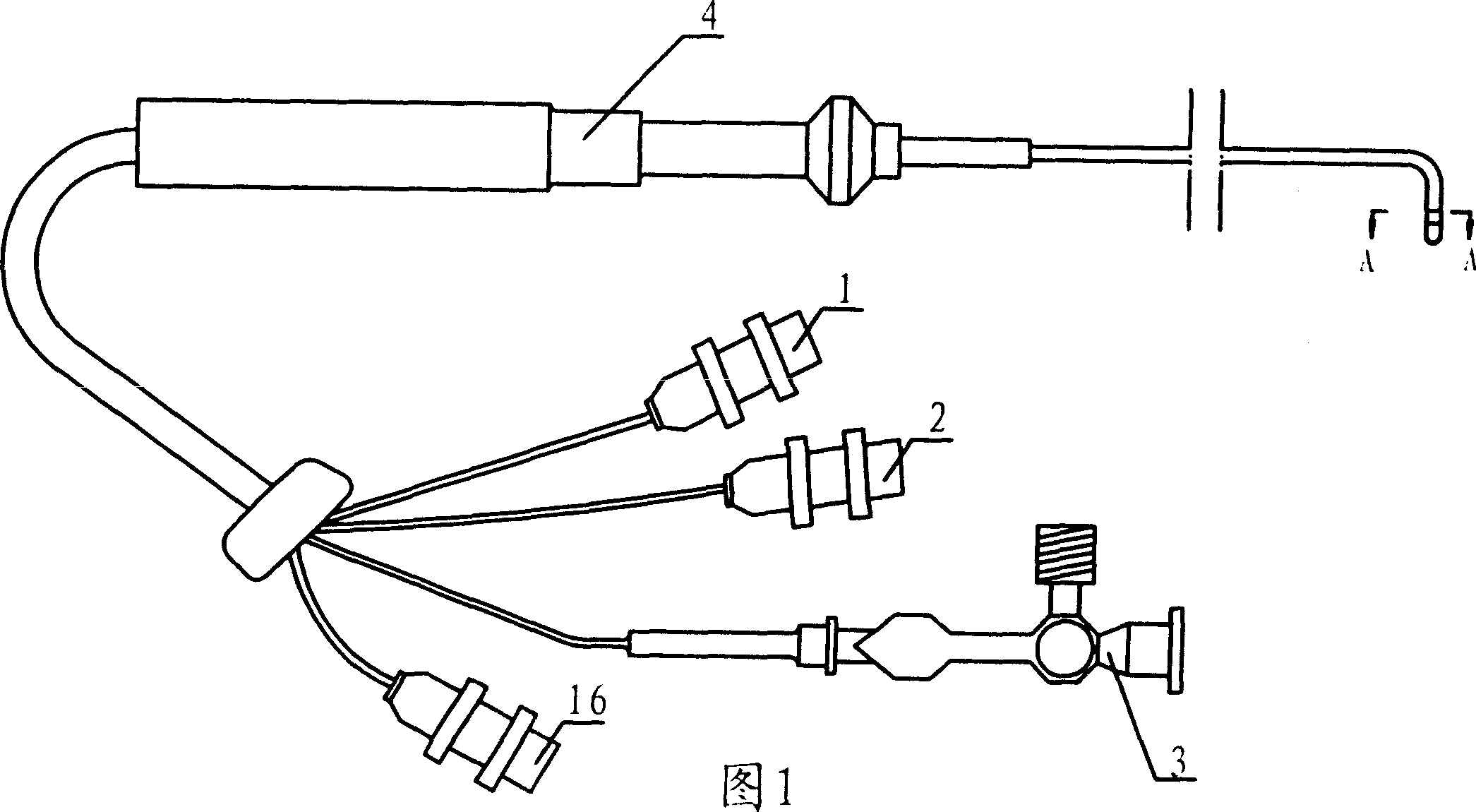
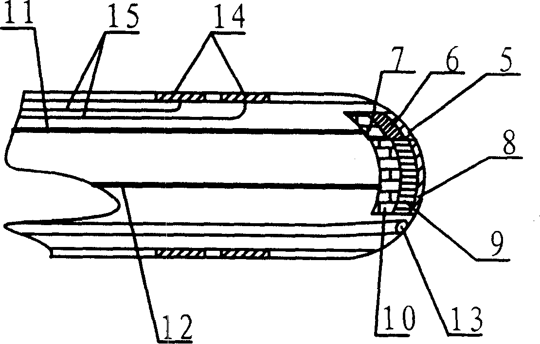
[0030] Set up a multifunctional ultrasonic catheter carried by the interventional catheter, and combine different types and numbers of transducers according to needs. According to different detection locations and purposes, the ultrasonic transducers installed on the top of the multifunctional ultrasonic catheter can be set to different sizes and shape, form different (ring, sector, rectangular, etc.) emitting surfaces and irradiation surfaces, extend the multifunctional ultrasound catheter to the detected part, and collect ultrasound image information and Doppler information at all levels of the target tissue, and the original radio frequency Information, etc., the acquired original information is input into the CPU of the host, and the corresponding analysis software and image processing system on the host are started according t...
Embodiment 2
[0032] Embodiment 2, the ultrasonic irradiation process method of entering the cardiovascular cavity comprises the following steps:
[0033] 1. Establish a multifunctional ultrasound catheter that meets the requirements of cardiovascular intervention
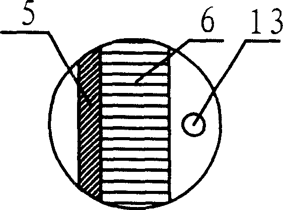
[0034] There are strict size requirements for entering the cardiovascular cavity. For example, the outer diameter of the multifunctional ultrasonic catheter entering the arterial system is required to be within 8F (2.67mm), generally within 7F (2.33mm), and can achieve 16 or more crystal array ultrasound imaging. The basic goal is that the above-mentioned crystal arrays are arranged in a small convex formation. There is a 0.4mm hole in the multifunctional ultrasonic catheter, which allows the 0.35mm guide wire and the 0.3mm micro-injection needle to pass through. The multi-functional ultrasound catheter head enters the detection ultrasound detection area behind. There is still a small area at the tip of the catheter for the pl...
Embodiment 3
[0043] Example 3: Diagnosis and Treatment Process of Ultrasonic Radiation into Other Viscera
[0044] (1) Establish the above-mentioned multifunctional ultrasound catheter. The size of the transducer on the catheter depends on the organ it enters. For example, the size of the catheter entering the bladder and renal pelvis is similar to the above-mentioned catheter entering the cardiovascular cavity, while entering the cavity such as the uterus For larger organs, the size of the multifunctional ultrasound catheter can be similar to the size of the head of the hysteroscope. The number, shape, and mode of emitting and obtaining ultrasound at the front end of the multifunctional ultrasound catheter can be changed according to the different organs and purposes.
[0045] (2) The operation process is the same as clinical cystoscopy and hysteroscopy, but a certain amount of normal saline should be injected in advance to make the multifunctional ultrasonic catheter immersed in water to ...
PUM
 Abstract
Description
Claims
Application Information
 - R&D Engineer
- R&D Manager
- IP Professional
- Industry Leading Data Capabilities
- Powerful AI technology
- Patent DNA Extraction
Browse by: Latest US Patents, China's latest patents, Technical Efficacy Thesaurus, Application Domain, Technology Topic, Popular Technical Reports.
© 2024 PatSnap. All rights reserved.Legal|Privacy policy|Modern Slavery Act Transparency Statement|Sitemap|About US| Contact US: help@patsnap.com


