Segmentation method and device for inner myocardial membrane and/or outer myocardial membrane of left ventricle of heart

A technology of epimyocardium and left ventricle, applied in the field of image processing, can solve problems such as segmentation deviation, low robustness, and strict requirements on segmentation accuracy, so as to reduce segmentation error, improve segmentation accuracy and robustness, reduce The effect of segmentation complexity

Active Publication Date: 2022-04-29
SHENZHEN KEYA MEDICAL TECH CORP
View PDF12 Cites 2 Cited by
  • Summary
  • Abstract
  • Description
  • Claims
  • Application Information

AI Technical Summary

Problems solved by technology

Due to the thin-layer structure of the left ventricular myocardium, the direct prediction of the myocardium is very demanding on the accuracy of the segmentation. Without the prio

Method used


Image

  • Segmentation method and device for inner myocardial membrane and/or outer myocardial membrane of left ventricle of heart
  • Segmentation method and device for inner myocardial membrane and/or outer myocardial membrane of left ventricle of heart
  • Segmentation method and device for inner myocardial membrane and/or outer myocardial membrane of left ventricle of heart

Examples


Embodiment Construction

[0019] In order to enable those skilled in the art to better understand the technical solutions of the present disclosure, the present disclosure will be described in detail below in conjunction with the accompanying drawings and specific embodiments. Embodiments of the present disclosure will be described in further detail below in conjunction with the accompanying drawings and specific embodiments, but are not intended to limit the present disclosure. For the various steps described herein, if there is no need for a contextual relationship between each other, the order described herein as an example should not be considered as a limitation, and those skilled in the art will know that the order can be adjusted, as long as It is enough not to destroy the logic between them so that the whole process cannot be realized.

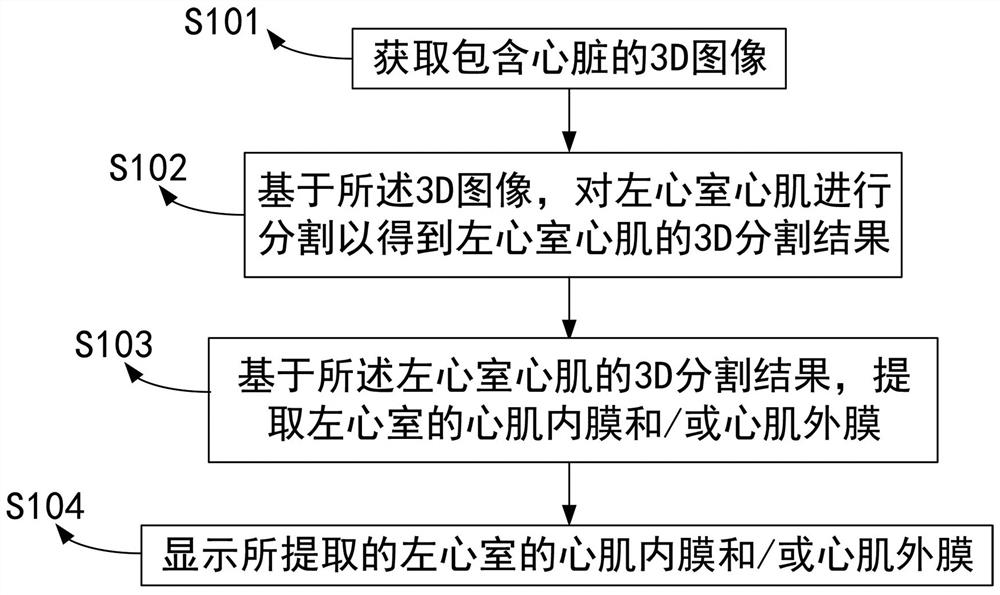
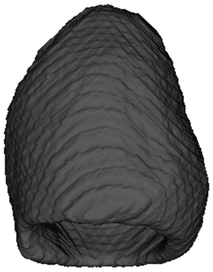
[0020] figure 1 A flow chart showing a method for segmenting the endocardium and / or epimyocardium of the left ventricle of the heart according to an embodimen...


PUM


Abstract

The invention provides a method for segmenting an inner myocardial membrane and/or an outer myocardial membrane of a left ventricle of a heart, and the method comprises the steps: obtaining a 3D image containing the heart through a processor; based on the 3D image, segmenting the left ventricular myocardium to obtain a 3D segmentation result of the left ventricular myocardium; based on the 3D segmentation result of the left ventricular myocardium, extracting an inner myocardial membrane and/or an outer myocardial membrane of the left ventricle; and displaying the extracted inner and/or outer myocardial membranes of the left ventricle. The method can improve the segmentation efficiency, reduce the segmentation error and improve the segmentation accuracy.

Description

technical field [0001] The present disclosure relates to the field of image processing, and more particularly, to a method and device for segmenting endocardium and / or epimyocardium of the left ventricle of the heart. Background technique [0002] Segmentation of left ventricular myocardium is an important step in cardiac function analysis. Functional evaluations such as left ventricular wall thickness and ejection fraction, and diagnosis and treatment of hypertrophic cardiomyopathy are all based on accurate segmentation of left ventricular myocardium. In the image of the left ventricle of the heart, since the gray values ​​of the left ventricular endocardium and papillary muscle, trabecula and other tissues are very close, and the gray value difference between the left ventricular epicocardium and the external adjacent tissues is small, the segmentation task has a large challenging. [0003] Based on traditional image processing methods, a 3D image is generally given and i...

Claims


Application Information

Patent Timeline
IPC IPC(8): G06T7/00G06T7/11G06T7/12G06T7/13G06N3/04G06N3/08
CPCG06T7/0012G06T7/11G06T7/12G06T7/13G06N3/084G06T2207/20081G06T2207/20084G06T2207/30048G06N3/045
Inventor 安木军李俊环丁志敏李育威曹坤琳王立伟
Owner SHENZHEN KEYA MEDICAL TECH CORP
Why Eureka
Try Eureka