Ultrasonic image quantitative diagnosis system and signal processing method thereof

An ultrasound image and image quantification technology, applied in ultrasound/sonic/infrasound image/data processing, image data processing, ultrasound/sonic/infrasonic Permian technology, etc. , to achieve the effect of improving image quality

Pending Publication Date: 2022-04-29
漯河市骨科医院(漯河市第五人民医院漯河医学高等专科学校第二附属医院)
View PDF0 Cites 0 Cited by
  • Summary
  • Abstract
  • Description
  • Claims
  • Application Information

AI Technical Summary

Problems solved by technology

However, different ultrasound image processing methods are not targeted for different organs, resulting in the inability to achieve optimal denoising ultrasound images of different organs

Method used


Image

  • Ultrasonic image quantitative diagnosis system and signal processing method thereof
  • Ultrasonic image quantitative diagnosis system and signal processing method thereof
  • Ultrasonic image quantitative diagnosis system and signal processing method thereof

Examples


Embodiment Construction

[0046] The following will clearly and completely describe the technical solutions in the embodiments of the present invention with reference to the accompanying drawings in the embodiments of the present invention. Obviously, the described embodiments are only some, not all, embodiments of the present invention. Based on the embodiments of the present invention, all other embodiments obtained by persons of ordinary skill in the art without making creative efforts belong to the protection scope of the present invention.

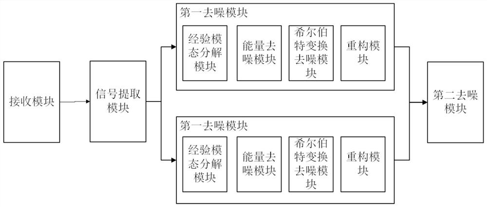
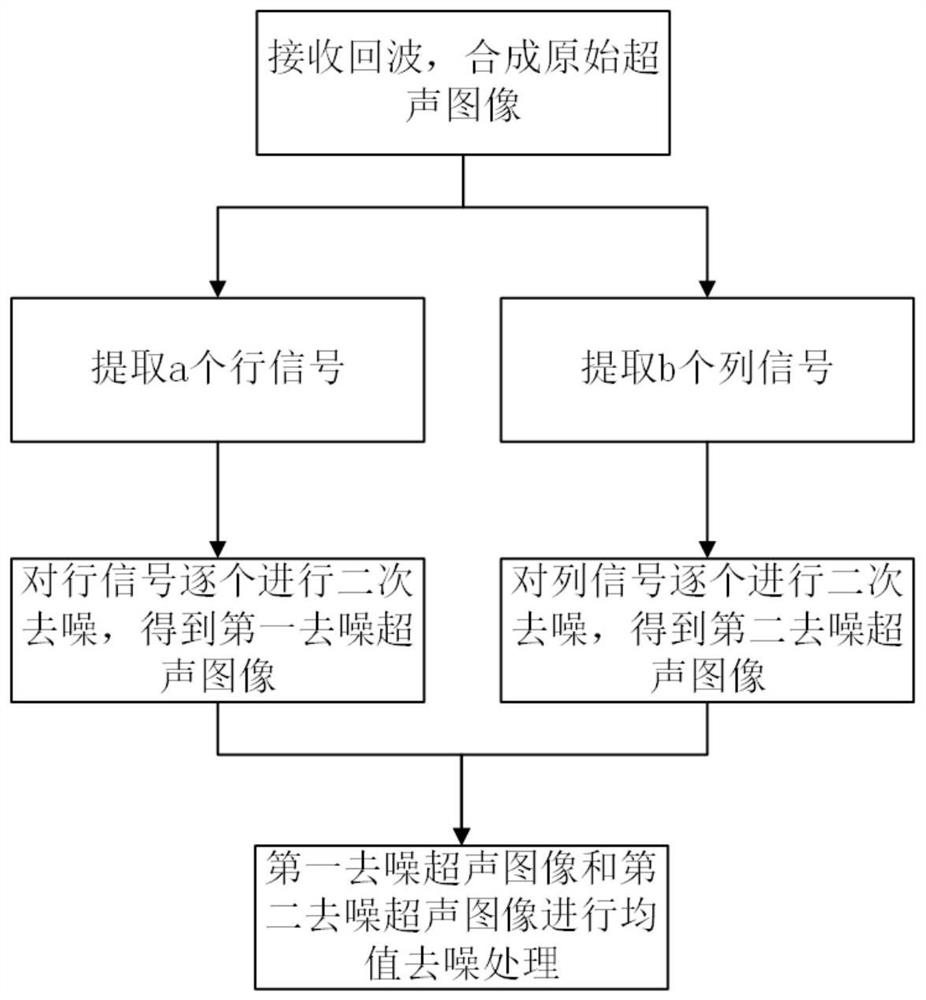
[0047] The present invention performs speckle suppression on ultrasound images of the liver. Since the liver is a substantial organ with hepatic lobules inside, its ultrasound noise distribution is different from that of kidneys, prostate and other organs.

[0048] figure 1 It is a structural diagram of the ultrasonic image quantification diagnosis system of the present invention. The present invention discloses an ultrasonic image quantitative diagnosis syst...


PUM


Abstract

The invention discloses an ultrasonic image quantitative diagnosis system and a signal processing method thereof. The ultrasonic image quantitative diagnosis system comprises a data receiving module, a first denoising module and a second denoising module, an empirical mode decomposition module in the first denoising module is used for carrying out empirical mode decomposition on each row of signals / column of signals of an ultrasonic image to obtain an eigenmode function and a margin, and then secondary denoising of the signals is achieved through an energy denoising method and a Hilbert transform denoising method. For speckle distribution of the liver ultrasonic image, the ultrasonic image is denoised through a row signal method and a column signal method, and noise residues in a certain direction occurring in denoising through a single method can be effectively improved by taking the mean value of the row signal method and the column signal method; and during signal processing, through an energy denoising method and a Hilbert denoising method, secondary denoising can further eliminate noise of a low-frequency signal after primary high-frequency denoising, so that the quality of a liver ultrasonic image is improved, and an accurate basis is provided for diagnosis of a doctor.

Description

technical field [0001] The invention belongs to the field of ultrasonic detection, and in particular relates to an ultrasonic image quantitative diagnosis system and a signal processing method thereof. Background technique [0002] Ultrasound imaging images are widely used in various medical institutions due to their low price, simple operation, and no radiation. Compared with CT and MRI, ultrasonic imaging technology has many advantages, but at the same time, it also has problems such as low image resolution and serious interference of speckle noise. This is because during ultrasound imaging, the biological tissue acts as a scattering cutoff during the imaging process, resulting in constructive and destructive interference, and speckles (speckles) with alternating light and dark appear in the acquired ultrasound image. [0003] Speckle is actually a phenomenon that occurs when an ultrasound image contains two types of noise, one is Gaussian noise that can be ignored, which...

Claims


Application Information

Patent Timeline
Patent Type & Authority Applications(China)
IPC IPC(8): G06T7/00G06K9/00A61B8/08
CPCG06T7/0012A61B8/5269G06T2207/10132G06T2207/30056G06F2218/10G06F2218/04
Inventor 庄艳芳杜效伟王惠娟朱红丽
Owner 漯河市骨科医院(漯河市第五人民医院漯河医学高等专科学校第二附属医院)
Why Eureka
Try Eureka