
Three-dimensional MRI brain tumor segmentation method based on deep learning

A deep learning and brain tumor technology, applied in neural learning methods, image analysis, image data processing, etc., can solve problems such as single feature scale, poor brain tumor segmentation effect, lack of multi-scale and global context information in semantic features, etc. Achieve the effect of reducing the influence of redundant features and improving the segmentation ability

Pending Publication Date: 2022-04-15
NANCHANG UNIV
View PDF0 Cites 4 Cited by
  • Summary
  • Abstract
  • Description
  • Claims
  • Application Information

AI Technical Summary

Problems solved by technology

However, all convolutional modules are composed of 2 stacked 3 convolutional layers, which leads to a relatively single scale of extracted features, and the lack of multi-scale and global context information in the captured semantic features, making it more challenging in brain tumors. poor segmentation

Method used


Image

  • Three-dimensional MRI brain tumor segmentation method based on deep learning
  • Three-dimensional MRI brain tumor segmentation method based on deep learning
  • Three-dimensional MRI brain tumor segmentation method based on deep learning

Examples


Embodiment Construction

[0026] The present invention will be further elaborated in conjunction with the accompanying drawings.

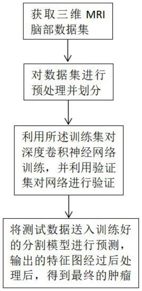
[0027] A 3D MRI brain tumor segmentation method based on deep learning, such as figure 1 shown, including the following steps:

[0028] S1. Preprocessing the 3D MRI brain data and dividing the data set to meet the input conditions of the model;

[0029] MRI images have 4 different modalities including T1, T1ce, T2, and FLAIR, and we splice the 4 data together to form 4 input channels. Usually, the proportion of background information in the whole image is relatively large, and the proportion of tumor area is very small, which will lead to serious data imbalance, and the background is not helpful for segmentation, so we choose to remove the background around the brain area Information, crop the 3D brain MRI image from the original size of 155*240*240 to the size of 150*192*192. The size of the 3D image finally sent to the network is 96*144*144.

[0030] In addition, data...


PUM


Abstract

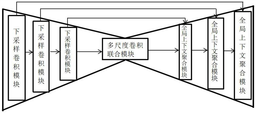
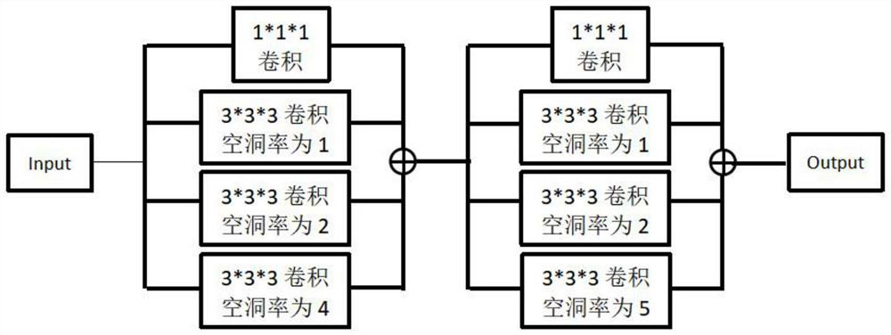
The invention discloses a three-dimensional MRI brain tumor segmentation method based on deep learning, and the method comprises the following steps: S1, carrying out the preprocessing of three-dimensional MRI brain data, dividing a data set, and enabling the data set to meet the input conditions of a model; s2, a deep convolutional neural network is constructed and trained, a network framework adopts the form of an encoder and a decoder, and a multi-scale convolution joint module and a global context aggregation module are added; and S3, post-processing the obtained prediction data to further improve the segmentation effect. According to the segmentation method provided by the invention, the low-level features and the high-level features of the segmentation object are combined, the multi-scale information and the global context information are effectively fused, and the influence of the learned redundant features is reduced, so that the segmentation result of the brain tumor is improved.

Description

technical field [0001] The invention relates to the field of medical image segmentation, in particular to a three-dimensional MRI brain tumor segmentation method based on deep learning. Background technique [0002] Predicting full 3D point clouds is a core task in many computer vision tasks. Brain tumors have a very high mortality and morbidity rate, but if brain tumors are detected in time, early diagnosis and early treatment can increase the possibility of cure. Brain tumor image segmentation is a very important step in the clinical diagnosis and treatment of brain tumors. By segmenting tumors in MRI images, doctors can locate the location of the tumor and obtain the size of the tumor, and then formulate relevant treatment and rehabilitation strategies. However, due to the characteristics of brain tumors such as complex structure, variable shape and extremely unbalanced categories, traditional image segmentation algorithms such as region growing and thresholding methods ...

Claims


Application Information

Patent Timeline
Patent Type & Authority Applications(China)
IPC IPC(8): G06T7/10G06N3/04G06N3/08
Inventor 邹艳妮王泽坤刘小平刘捷
Owner NANCHANG UNIV
Learn More