Brain tumor MRI image three-dimensional segmentation method based on RAPNet network

A brain tumor and network technology, applied in the field of image processing, can solve the problem of low segmentation accuracy

Pending Publication Date: 2022-03-18
CHONGQING UNIV OF POSTS & TELECOMM
View PDF0 Cites 3 Cited by
  • Summary
  • Abstract
  • Description
  • Claims
  • Application Information

AI Technical Summary

Problems solved by technology

[0006] In order to solve the problem that brain tumors present different sizes and shapes in MRI images of different patients, resulting in low segmentation accuracy based on single-scale DCNN, the present invention proposes a three-dimensional segmentation method for brain tumor MRI images based on RAPNet network.

Method used


Image

  • Brain tumor MRI image three-dimensional segmentation method based on RAPNet network
  • Brain tumor MRI image three-dimensional segmentation method based on RAPNet network
  • Brain tumor MRI image three-dimensional segmentation method based on RAPNet network

Examples


Embodiment Construction

[0055] Next, the technical solutions in the embodiments of the present invention will be described in connection with the drawings of the embodiments of the present invention, and it is understood that the described embodiments are merely the embodiments of the present invention, not all of the embodiments. Based on the embodiments of the present invention, all other embodiments obtained by those of ordinary skill in the art are in the range of the present invention without making creative labor premise.

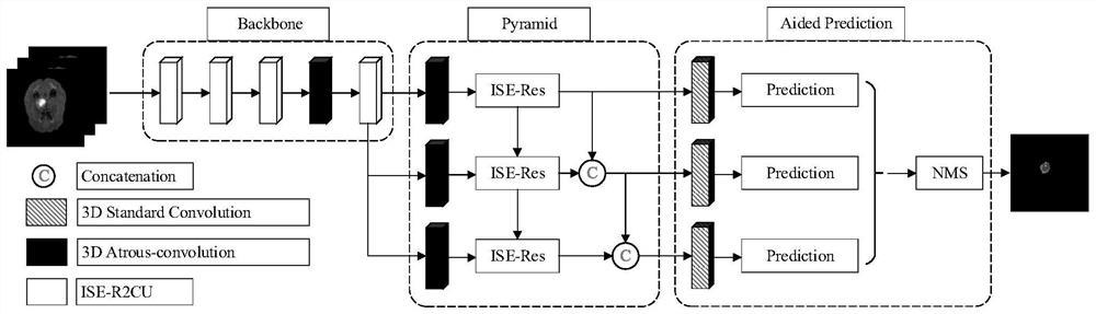
[0056] The present invention provides a three-dimensional division method based on a brain tumor MRI image based on a RAPNET network, comprising the steps of:

[0057] Build a RAPNET network and train them;

[0058] The brain MRI image will be input into the training-well-trained RAPNET network to perform image recognition segmentation, resulting in divided brain tumor MRI images and its sub-structural area;

[0059] The RAPNET network includes a backbone network, a feature pyra...


PUM


Abstract

The invention belongs to the field of image processing, and particularly relates to a brain tumor MRI image three-dimensional segmentation method based on an RAPNet network, and the method comprises the steps: constructing the RAPNet network, and carrying out the training of the RAPNet network; the brain MRI image is input into a trained RAPNet network for image recognition and segmentation, a segmented brain tumor MRI image and a substructure region thereof are obtained, the RAPNet network comprises a backbone network, a feature pyramid and auxiliary prediction, and the backbone network is composed of a cavity convolution and a plurality of ISE-R2CU units and used for extracting shallow features and deep features of the input image; according to the method, the feature pyramid formed by the 3D cavity convolution and the cross-model attention mechanism is combined with the trunk to learn the effective features of the whole tumor and the substructure thereof, so that the method has the advantage of fitting various tissue boundaries in the tumor.

Description

Technical field [0001] The present invention belongs to the field of image processing, and specifically, a three-dimensional division method based on a cerebral tumor MRI image based on a RAPNET network. Background technique [0002] Brain tumors are abnormal cells aggregated in the brain, which is an extremely dangerous disease. Tumor cells will be rapidly divided and infinitely proliferate, and the human central nervous system will eventually lead to death. Medical images provide technical support for early diagnosis of brain tumors. In numerous imaging methods, magnetic resonance imaging, mri technology provides superior contrast for multi-directional imaging of brain soft tissue, and has no invasive, radiated radiation. The characteristics, so MRI has become a common technique for diagnosing cerebral tumors. During treatment, the segmentation of tumor tumors is especially important before the treatment of normal cells while killing tumor cells. However, using human methods to...

Claims


Application Information

Patent Timeline
Patent Type & Authority Applications(China)
IPC IPC(8): G06T7/11G06N3/04G06N3/08
CPCG06T7/11G06N3/084G06T2207/10028G06T2207/10088G06T2207/30016G06T2207/30096G06T2207/20081G06T2207/20084G06T2207/20221G06N3/047G06N3/048G06N3/045
Inventor 胡敏熊思黄宏程
Owner CHONGQING UNIV OF POSTS & TELECOMM
Why Eureka
Try Eureka