Anterior segment OCT (optical coherence tomography) imaging system for ophthalmic examination

An imaging system and ophthalmic examination technology, applied in application, medical science, equipment for testing eyes, etc., can solve the problems of insufficient detail and comprehensiveness of the OCT imaging analysis system, and achieve the effect of improving the detail and comprehensiveness.

Inactive Publication Date: 2022-03-01
THE FIRST AFFILIATED HOSPITAL OF ZHENGZHOU UNIV
View PDF0 Cites 0 Cited by
  • Summary
  • Abstract
  • Description
  • Claims
  • Application Information

AI Technical Summary

Problems solved by technology

[0004] Aiming at the deficiencies in the prior art, the purpose of the present invention is to provide an anterior segment OCT imaging system for ophthalmological examination, which can independently analyze ocular trauma, and obtain special signals in the detection process, improving the meticulousness of detection and analysis , to solve the problem that the existing OCT imaging analysis system is not detailed and comprehensive enough

Method used


Image

  • Anterior segment OCT (optical coherence tomography) imaging system for ophthalmic examination
  • Anterior segment OCT (optical coherence tomography) imaging system for ophthalmic examination
  • Anterior segment OCT (optical coherence tomography) imaging system for ophthalmic examination

Examples


Embodiment 1

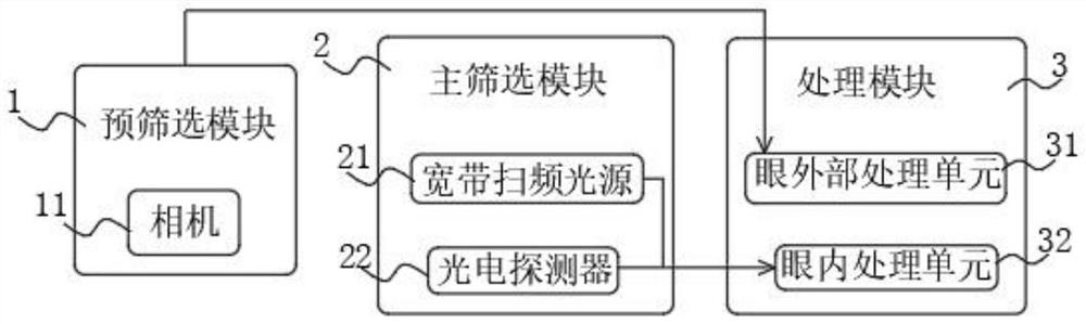
[0034] Example 1, please refer to figure 1 , an anterior segment OCT imaging system for ophthalmic examination, the imaging system includes: a pre-screening module 1 , a main screening module 2 and a processing module 3 .

[0035] The pre-screening module 1 is used to acquire the external image of the eye; the pre-screened module 1 includes a camera 11, and the camera 11 is used to acquire the external image of the eye and the contrast area image.

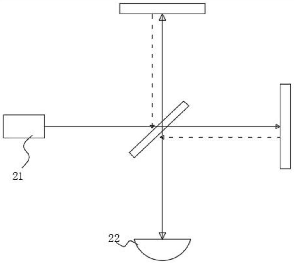
[0036] see image 3 , the main screening module 2 is used to obtain the OCT image of the anterior segment of the eyeball; the main screening module 2 includes a broadband sweeping light source 21 and a photodetector 22, and the broadband sweeping light source 21 is used to emit a sweeping spectrum signal, The photodetector 22 is used to record frequency-swept spectral signals.

[0037] The processing module 3 is used to process the acquired external ocular image and the OCT imaging of the anterior segment of the eye to obtain cor...

Embodiment 2

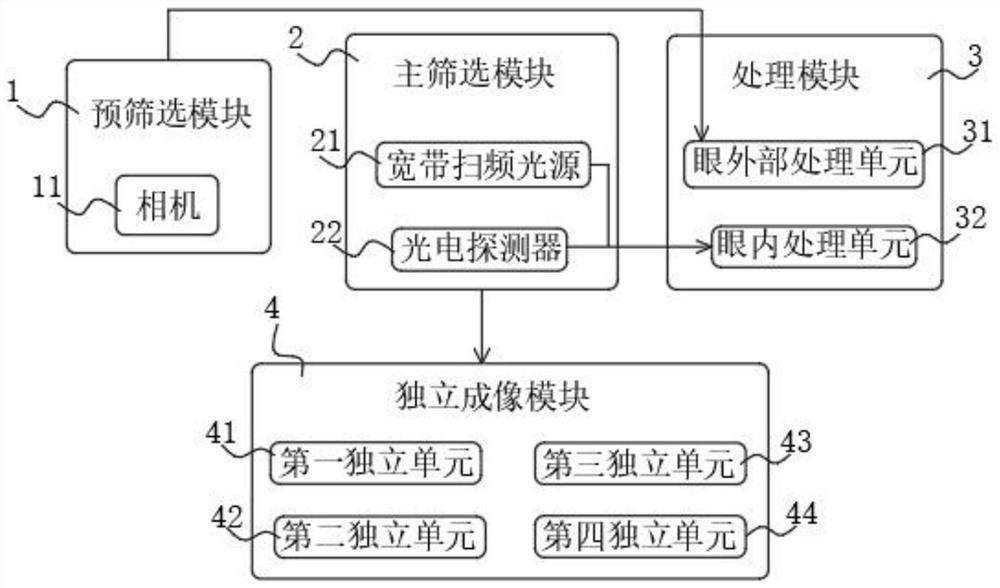
[0049] Example 2, please refer to figure 2 In the second embodiment, an independent imaging module 4 is added on the basis of the imaging obtained in the first embodiment, and the independent imaging module 4 can be used for independent imaging analysis of eye detection, which improves the meticulousness of the independent imaging analysis. The imaging system also includes an independent imaging module 4, the main screening module 2 is used to obtain the OCT image of the anterior segment of the eyeball; the independent imaging module 4 is used to independently decompose the OCT image of the anterior segment of the eyeball, and obtain anterior segment of the eyeball OCT images of different parts.

[0050] The independent imaging module 4 includes a first independent unit 41, a second independent unit 42, a third independent unit 43 and a fourth independent unit 44, the first independent unit 41 is used to obtain the imaging image of the cornea, and the second independent unit ...


PUM


Abstract

The invention provides an anterior segment OCT (optical coherence tomography) imaging system for ophthalmic examination. The imaging system comprises a pre-screening module, a main screening module, a processing module and an independent imaging module, the pre-screening module is used for acquiring an eye external image; the main screening module is used for acquiring an OCT image of an anterior segment of an eyeball; the processing module is used for processing the acquired external eye image and the OCT image of the anterior segment of the eyeball to obtain a corresponding imaging result; the independent imaging module is used for independently decomposing the OCT image of the anterior segment of the eyeball and obtaining OCT images of different parts of the anterior segment of the eyeball, the eye trauma condition can be independently analyzed, special signals in the detection process are obtained, the meticulous performance of detection analysis is improved, and the detection accuracy is improved. The problem that an existing OCT imaging analysis system is not meticulous and comprehensive enough is solved.

Description

technical field [0001] The invention relates to the technical field of ophthalmology detection, in particular to an anterior segment OCT imaging system for ophthalmology inspection. Background technique [0002] The anterior segment, also known as the anterior segment, refers to the part located in front of the lens, specifically including: anterior chamber, posterior chamber, lens suspensory ligament, chamber angle, part of the lens, peripheral vitreous body, retina and extraocular muscle attachment points and conjunctiva, etc., the anterior segment Clinical signs of various disorders, including eyelid and adnexa, conjunctivopathy, corneal disease, sarcoid and pupillary abnormalities, glaucoma, cataracts, strabismus, orbitopathy and exophthalmos, ocular trauma, and ocular syndromes. Optical coherence tomography (OCT) is a new three-dimensional tomography technology developed gradually in the 1990s. OCT is based on the principle of low-coherence interference to obtain tomog...

Claims


Application Information

Patent Timeline
Patent Type & Authority Applications(China)
IPC IPC(8): A61B3/10A61B3/14
CPCA61B3/102A61B3/14A61B3/0016
Inventor 赵红
Owner THE FIRST AFFILIATED HOSPITAL OF ZHENGZHOU UNIV
Why Eureka
Try Eureka