


Quick Research
Generate reliable direction feasibility study reports for your R&D in just a few steps.

Technical Q&A
Discover and master advanced knowledge NOW. Basics, ideas, possibilities, all at once.

Find Solutions
As an expert in R&D theories, this can generate solutions to your technical problems instantly.

Evaluate Feasibility
Analyze your overall solution with one click, know your potential R&D risks in advance.

Monitor Landscape
Get weekly tech updates, stay abreast of the latest tech innovations and key insights.
Biomarker analysis method based on multiple immunohistochemical technologies and application thereof
A biomarker and immunohistochemical technology, applied in the field of biological detection and analysis, can solve problems such as the inability to explain the spatial relationship of tumor cells well, and achieve the effect of reducing complicated work.
- Summary
- Abstract
- Description
- Claims
- Application Information
AI Technical Summary
Problems solved by technology
Method used
Image

Examples
Embodiment 1
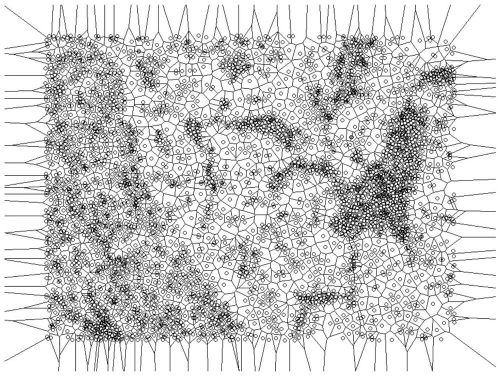
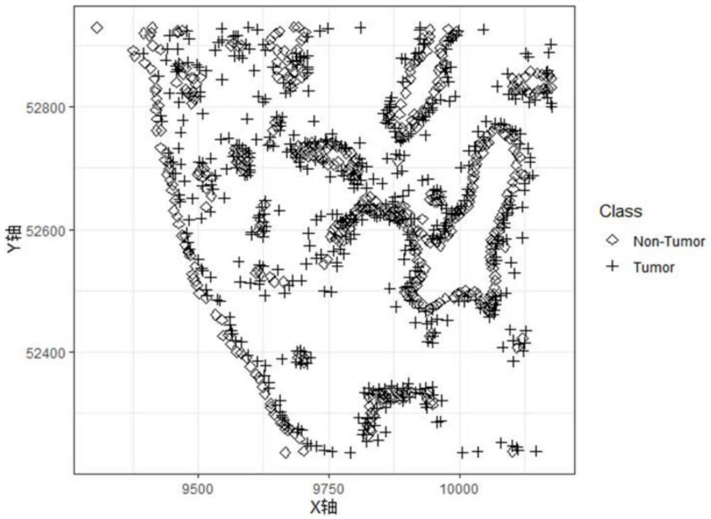
[0075] Sample used in this embodiment is a clinical patient (Patient1) tumor tissue sample 1 case of lung cancer in laboratory testing. 7 the completed multi-color MOTiF immunohistochemical staining of tissue samples of tumor patients (e.g., for lung cancer TM PD-1 / PD-L1 Panel kit, comprising a targeting FoxP3, PD-L1, PanCK, PD-1, CD8, 6 antibodies for CD68), for the labeling fluorescent-positive cells, and through the entire film system VECTRA POLARIS Get scanning microscopy immunohistochemistry panorama. Phenochart by selecting the target area (tumor inner cells containing, the boundary region of the tumor cells and stromal cells), introduced inForm system, using official lung algorithm predefined rule-based classification acquired morphological and phenotypic changes, export data for biological marker analysis and calculation.
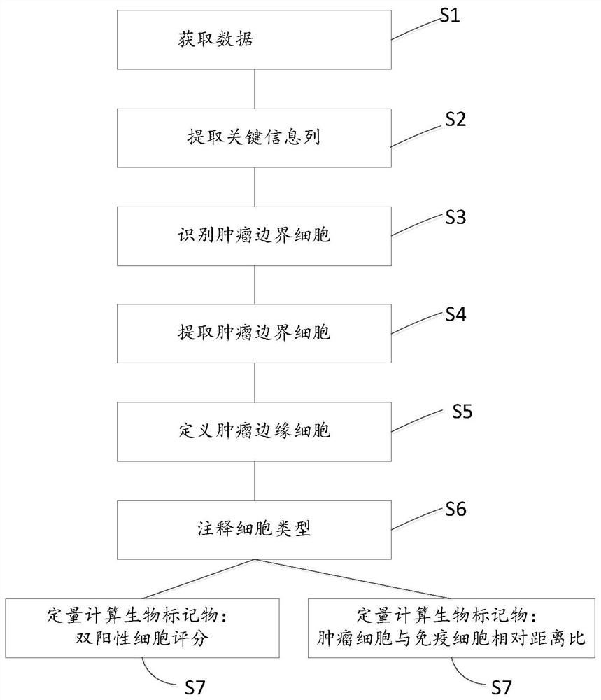
[0076] Biomarker analysis following steps:
[0077] 1. Read Data: Import "* cell_seg_data.txt" data to RStudio;
[0078] 2. Data extraction: extract ...
Embodiment 2
[0089] Sample used in this embodiment is (Patient2) tumor tissue sample in another one embodiment the clinical laboratory testing of lung cancer patients. Completed the patient tissue tumor sample multiple immunohistochemical staining (color 6 7 standard: FoxP3, PD-L1, PanCK, PD-1, CD8, CD68), and after VECTRA POLARIS slide scanning system for full acquisition Immunohistochemistry microscopic panorama. Phenochart by selecting the target area (tumor inner cells containing, the boundary region of the tumor cells and stromal cells), introduced into the system inForm, lung using a predefined algorithm official rules are organized based on morphological classification and typing of cells derived biological data for marker analysis and calculation.
[0090] Biomarker analysis following steps:
[0091] 1. Read Data: Import "* cell_seg_data.txt" data to RStudio;
[0092]2. Extract data: Extract critical information columns (including: organizational morphological classification columns, c...
PUM
 Abstract
Description
Claims
Application Information
 - R&D Engineer
- R&D Manager
- IP Professional
- Industry Leading Data Capabilities
- Powerful AI technology
- Patent DNA Extraction
Browse by: Latest US Patents, China's latest patents, Technical Efficacy Thesaurus, Application Domain, Technology Topic, Popular Technical Reports.
© 2024 PatSnap. All rights reserved.Legal|Privacy policy|Modern Slavery Act Transparency Statement|Sitemap|About US| Contact US: help@patsnap.com