Head positioning support for magnetic resonance photographing of temporomandibular joint and use method of head positioning support

A temporomandibular joint and positioning bracket technology, applied in the field of medical auxiliary equipment, can solve problems such as unusable and difficult measurement of related indicators

Pending Publication Date: 2021-09-03
SHANGHAI NINTH PEOPLES HOSPITAL SHANGHAI JIAO TONG UNIV SCHOOL OF MEDICINE
View PDF0 Cites 0 Cited by
  • Summary
  • Abstract
  • Description
  • Claims
  • Application Information

AI Technical Summary

Problems solved by technology

However, due to the lack of a unified reference plane when taking MRI images, there are large differences in the shooting angles and directions of several MRI images taken by different people or the same person before and after, which brings difficulties to the measurement of related indicators.
[0004] The X-ray cephalogram shooting bracket is fixed on the X-ray location film shooting machine and cannot be used during MRI shooting

Method used


Image

  • Head positioning support for magnetic resonance photographing of temporomandibular joint and use method of head positioning support

Examples


Embodiment Construction

[0024] The structural features of the present invention will be further elaborated below in conjunction with specific embodiments.

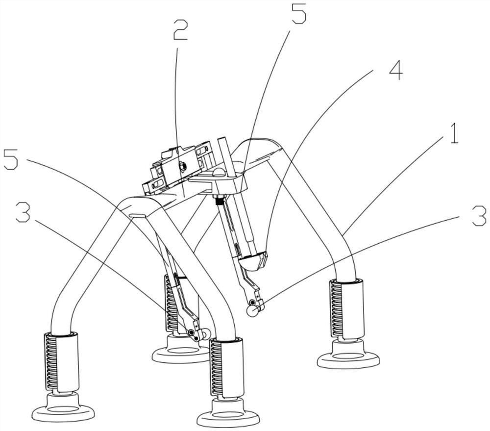
[0025] Such as figure 1 The shown head positioning bracket for magnetic resonance imaging of the temporomandibular joint includes a support frame placed on the magnetic resonance equipment, and the support frame has at least one beam 2; it also has a pair of ear positioning components 3 and a nose positioning part 4; the two ear positioning parts 3 are respectively positioned at the position of the ear when the human body is lying flat; the nose positioning part 4 is positioned at the corresponding position of the nose when the human body is lying flat ; The ear positioning part 3 and the nose positioning part 4 are respectively connected to the beam 2 through the adjustment assembly 5 .

[0026] The adjustment assembly 5 is a three-axis adjustment assembly or a universal adjustment assembly. As a cost-saving structure, the adjustment assembly ...


PUM


Abstract

The invention discloses a head positioning support for magnetic resonance photographing of a temporomandibular joint and a use method of the head positioning support. The head positioning support comprises a supporting frame arranged on a magnetic resonance device; the supporting frame is at least provided with a cross beam; a pair of ear positioning parts and a nose positioning part are further arranged; the two ear positioning parts are respectively positioned at positions of both ears when a human body lies flat; the nose positioning part is positioned at the position corresponding to the nose when the human body lies flat; and the ear positioning parts and the nose positioning part are connected with the cross beam through adjusting assemblies. The technical scheme of the invention has the following technical effects: a frankfort horizontal plane and the horizontal plane are kept consistent when different people or a same person is subjected to multiple times of photographing, so that subsequent measurement and comparison of relevant indexes (condylar process height, angle and the like) of the temporomandibular joint are facilitated, and a unified standard is established for magnetic resonance photographing and measurement of the temporomandibular joint.

Description

technical field [0001] The invention relates to a head fixing bracket, in particular to a head positioning bracket for magnetic resonance imaging of temporomandibular joints and a method for using the same, belonging to the technical field of medical auxiliary equipment. Background technique [0002] Temporomandibular joint disease is a relatively high incidence of clinical diseases, including temporomandibular joint disorder syndrome, temporomandibular joint ankylosis, temporomandibular joint tumor and other disease types. Because of its good soft tissue resolution, magnetic resonance can clearly show the structure and positional relationship between the articular disc and surrounding soft and hard tissues, so magnetic resonance examination of the temporomandibular joint is often necessary for such patients. [0003] At present, after the magnetic resonance imaging of the temporomandibular joint, in order to facilitate disease diagnosis and follow-up treatment, it is usuall...

Claims


Application Information

Patent Timeline
Patent Type & Authority Applications(China)
IPC IPC(8): A61B5/055
CPCA61B5/055A61B5/704
Inventor 陈敏洁谢千阳于洋刘芷扬杨驰董敏俊郑吉驷霍亮
Owner SHANGHAI NINTH PEOPLES HOSPITAL SHANGHAI JIAO TONG UNIV SCHOOL OF MEDICINE
Why Eureka
Try Eureka