
Ultrasonic three-dimensional imaging scanning device

A stereoscopic imaging and scanning device technology, applied in the directions of ultrasonic/sonic/infrasonic image/data processing, ultrasonic/sonic/infrasonic diagnosis, ultrasonic/sonic/infrasonic Permian technology, etc., capable of solving three-dimensional imaging, ultrasonic three-dimensional imaging Problems such as fuzzy computer simulation diagrams and synthetic images are blurred to achieve the effect of simple and convenient use, avoiding ionizing radiation damage, and accurate and convenient positioning

Pending Publication Date: 2021-06-25
SOOCHOW UNIV AFFILIATED CHILDRENS HOSPITAL
View PDF4 Cites 1 Cited by
  • Summary
  • Abstract
  • Description
  • Claims
  • Application Information

AI Technical Summary

Problems solved by technology

Due to the limitations of the current technology and the shortcomings of the detection method, the current synthetic image is blurred and cannot be clearly imaged, and cannot meet the clinical requirements such as three-dimensional restoration imaging of small blood vessels.
The current three-dimensional ultrasound imaging method is out of touch with clinical needs and cannot meet the clinical needs for image clarity and fineness
[0003] At present, there are ultrasonic three-dimensional imaging and ultrasonic four-dimensional technology, but the current imaging method has defects, and cannot provide clear and fast three-dimensional imaging. There are many limitations in the actual application process. The defects of the existing technology are:
[0004] 1. Ultrasound 3D imaging is fuzzy;
[0005] 2. Ultrasound 3D image restoration method is outdated, and the calculation is complicated and slow;
[0006] 3. Partial images can only be synthesized for specific layers of individual tissues, such as 4D echocardiography, which is only a 3D imaging of a certain layer corresponding to the cardiac cycle; 3D ultrasound imaging of the fetal face of a pregnant woman is only a fuzzy computer simulation
Because the abdomen of pregnant women is often not a regular spherical shape, and when the ultrasound probe is attached to the abdominal skin for ultrasound examination, there is a slight depression at the contact part of the probe and the skin

Method used


Image

  • Ultrasonic three-dimensional imaging scanning device

Examples


Embodiment Construction

[0019] The following will clearly and completely describe the technical solutions in the embodiments of the present invention with reference to the accompanying drawings in the embodiments of the present invention. Obviously, the described embodiments are only some, not all, embodiments of the present invention.

[0020] In describing the present invention, it should be understood that the terms "upper", "lower", "front", "rear", "left", "right", "top", "bottom", "inner", " The orientation or positional relationship indicated by "outside", etc. is based on the orientation or positional relationship shown in the drawings, and is only for the convenience of describing the present invention and simplifying the description, rather than indicating or implying that the referred device or element must have a specific orientation, so as to Specific orientation configurations and operations, therefore, are not to be construed as limitations on the invention.

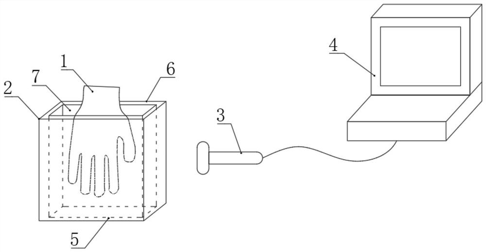
[0021] refer to figure 1...


PUM


Abstract

The invention discloses an ultrasonic three-dimensional imaging scanning device, and belongs to the field of medical ultrasonic three-dimensional imaging. The device comprises a scanning box, a cavity is formed in the scanning box, an opening is formed in the top of the scanning box, the cavity is filled with a coupling agent, an ultrasonic probe is arranged on the front side face or the rear side face of the scanning box, and the ultrasonic probe is arranged on the front side face or the rear side face of the scanning box; and the ultrasonic probe is electrically connected with the host. Three-dimensional imaging of blood vessels of the tablet hand is achieved through ultrasonic examination, and compared with invasive blood vessel examination, trauma of a patient is avoided, and ray ionizing radiation injury of the patient is avoided. Compared with traditional pure ultrasonic two-dimensional examination, the blood vessel three-dimensional deformation is restored more specifically and more vividly, positioning is more accurate and more convenient, and the using process is simple and convenient.

Description

technical field [0001] The invention relates to the technical field of medical ultrasonic stereo imaging, in particular to an ultrasonic stereo imaging scanning device. Background technique [0002] The current ultrasonic three-dimensional imaging or ultrasonic four-dimensional dynamic imaging is based on the synthesis of three-dimensional space images after two-dimensional ultrasonic examination, and at the same time, the changes of the images in a certain period of time are added to form ultrasonic four-dimensional dynamic imaging. Due to the limitations of the current technology and the shortcomings of the detection method, the current composite image is blurred and cannot be clearly imaged, and cannot meet the clinical requirements for three-dimensional restoration imaging of small blood vessels. The current three-dimensional ultrasound imaging method is out of touch with clinical needs and cannot meet the clinical needs for image clarity and fineness. [0003] At prese...

Claims


Application Information

Patent Timeline
Patent Type & Authority Applications(China)
IPC IPC(8): A61B8/00A61B8/08
CPCA61B8/0891A61B8/44A61B8/4444A61B8/466A61B8/483
Inventor 姚峰丁粤粤沈小芳朱兆峰文军印春花徐秋琴甄允方王晓东
Owner SOOCHOW UNIV AFFILIATED CHILDRENS HOSPITAL
Learn More