
Design method and application of probe combination for cancer detection

A cancer detection and design method technology, applied in the biological field, can solve the problem of inability to perform MRD monitoring, and achieve high sensitivity, specificity, and excellent coverage.

Active Publication Date: 2021-06-11
北京吉因加医学检验实验室有限公司 +1
View PDF9 Cites 4 Cited by
  • Summary
  • Abstract
  • Description
  • Claims
  • Application Information

AI Technical Summary

Problems solved by technology

For patients without or unable to provide tissue samples, this method cannot be used for MRD monitoring
(3) TAT and cost

Method used


Image

  • Design method and application of probe combination for cancer detection
  • Design method and application of probe combination for cancer detection
  • Design method and application of probe combination for cancer detection

Examples


Embodiment 1

[0056] Example 1 Probe combination and its target

[0057] 1.1 Design method and design results of capture probes

[0058] 1.1.1 Design method of capture probe

[0059] This example provides a cost-effective method for designing probe combinations that can be used for pan-cancer assistance, as follows:

[0060] (1) Determine the target cancer type, such as lung cancer, breast cancer, colorectal cancer, liver cancer, pancreatic cancer, gastric cancer, esophageal cancer, and bladder cancer.

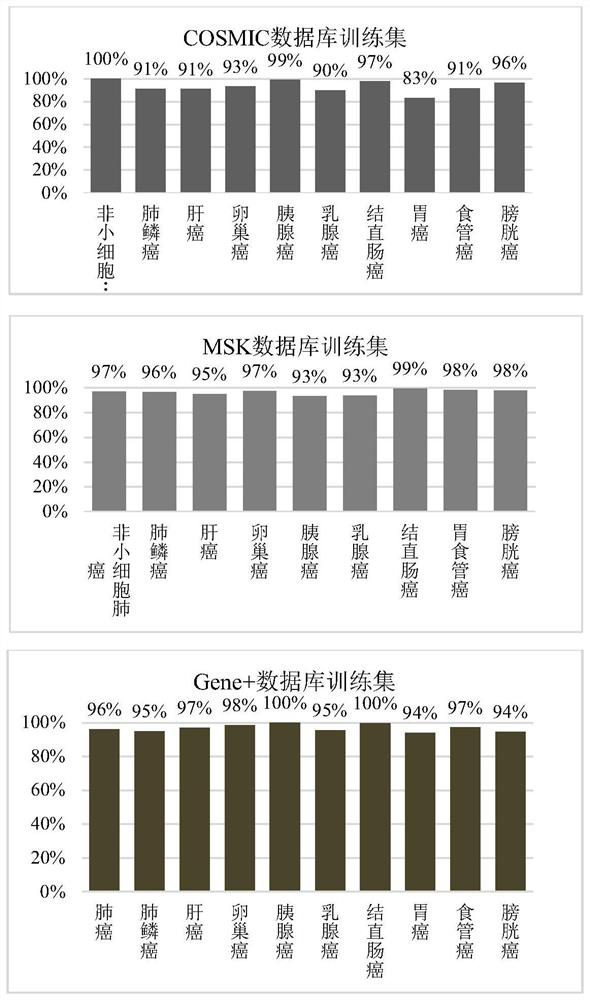
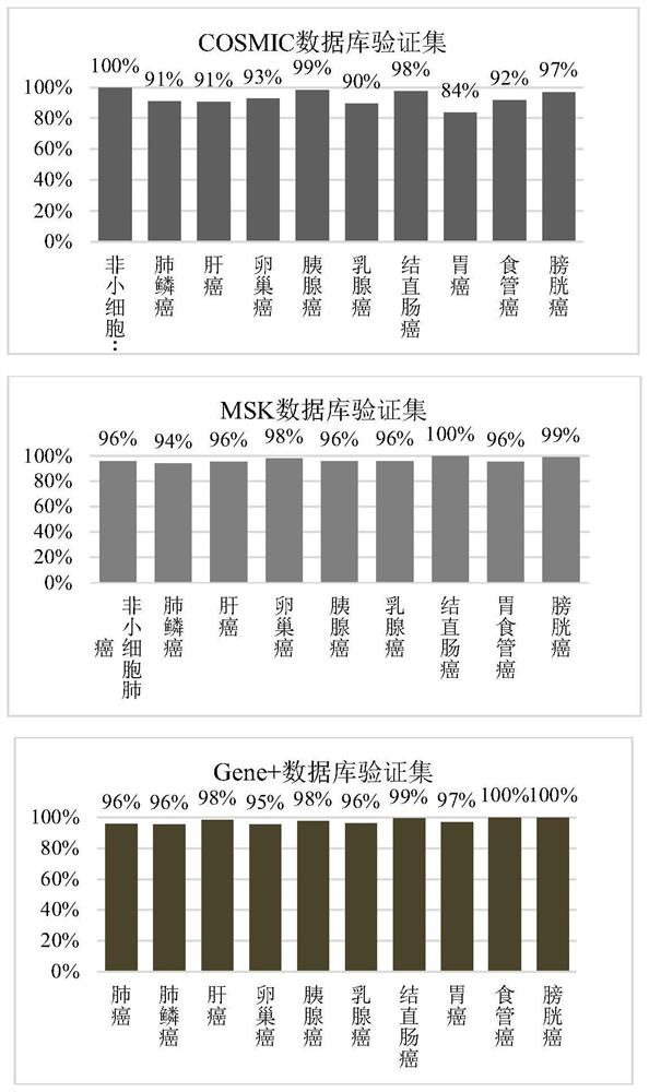
[0061] (2) Extract the mutation sets of nine major cancers in Gene+ database, COSMIC database and MSKCC database, and divide them into training set and verification set. Find the hotspot mutation area, merge the mutations whose reference genome distance is Muts . The formula is as follows:

[0062]

[0063] Among them, Reg Muts is the number of mutations in the merged region, Reg Len For the length of the merged region coverage interval, the merged region is expanded left and right...

Embodiment 2

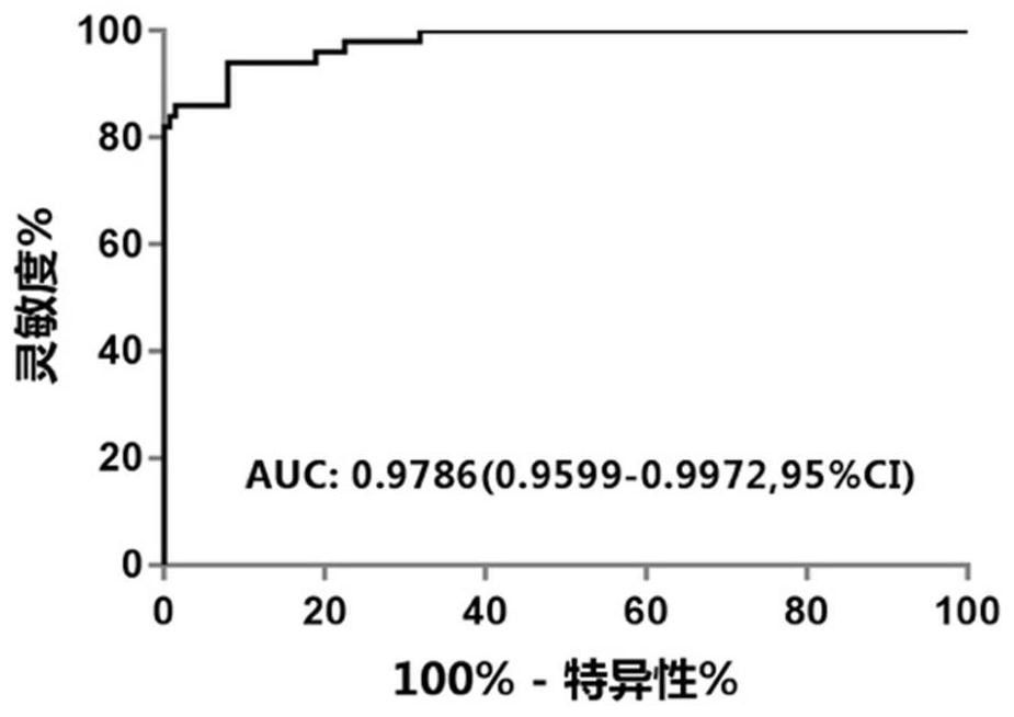
[0084] Example 2 DX testing application and sensitivity and specificity of early detection of liver cancer

[0085] Patients with stage I-III liver cancer without surgery and neoadjuvant therapy were recruited; at the same time, 200 healthy people without cancer history were recruited as a control group. Collect peripheral blood samples 10mL.

[0086] 2.1 Plasma separation and DNA extraction

[0087] For whole blood, plasma / blood cell separation should be performed in time (EDTA anticoagulant tube, within 4 hours; Streck tube within 72 hours), the separation steps are as follows:

[0088] (1) Centrifuge at 1600g for 10min at 4°C. After centrifugation, divide the upper layer of plasma into multiple 1.5mL or 2.0mL centrifuge tubes. Be careful not to absorb the white blood cells in the middle layer during the process of absorbing plasma.

[0089] After the plasma is separated in this step, the blood cells in the middle layer + bottom layer are reserved for use as a normal contr...

Embodiment 3

[0157] Example 3 Ovarian cancer, pancreatic cancer, early detection of colorectal cancer

[0158] Patients with stage I-III ovarian cancer, colorectal cancer, lung squamous cell carcinoma, and pancreatic cancer without surgery and neoadjuvant therapy were recruited to implement the test.

[0159] The detection method is the same as in Example 2.

[0160] Using the detection method of this project, the sensitivities of 36 cases of ovarian cancer, 79 cases of colorectal cancer, 28 cases of lung squamous cell carcinoma, and 35 cases of pancreatic cancer were 72%, 77%, 79% and 77%, respectively.


PUM


Abstract

The invention relates to a design method and application of a probe combination for cancer detection. The design method comprises the following steps: extracting a mutation set of cancer in a database, dividing the mutation set into a training set and a verification set, and combining mutations with reference genome distance less than or equal to 80 in the training set so as to obtain a plurality of mutational hotspot intervals; and sequentially screening the multiple mutation hotspot intervals on the basis of the regional mutation density, and taking the mutation hotspot intervals meeting the following conditions as targets of the probe combination. The probe combination designed by the invention has excellent coverage on common cancers, a Gene+ database and MSK database verification set is adopted to simulate the coverage condition of the panel on nine cancers, and the result shows that the coverage degrees of nine cancer types are all greater than 93%; the early cancer detection based on the probe has high sensitivity and specificity, and the detection rate of liver cancer reaches 85%; and a ctDNA positive judgment method based on the probe can effectively perform prognosis layering on a patient.

Description

technical field [0001] The invention relates to the field of biotechnology, in particular to a design method and application of a probe combination for cancer detection. Background technique [0002] Liver cancer is a type of malignant tumor with high incidence, and patients with liver cancer usually have a high mortality rate. The key reason for the high morbidity and mortality is the lack of effective early screening markers. In traditional diagnostic methods, serum alpha-fetoprotein (AFP) and liver ultrasonography are the main means for early screening of liver cancer, but related studies have shown that about 80% of small liver cancer patients (early liver cancer patients) have no significant AFP levels. increased, indicating that AFP has certain limitations as a screening marker and is not suitable for the detection of early liver cancer; and for early liver cancer, the sensitivity of ultrasound examination is only 47%; the sensitivity of detection by ultrasound combin...

Claims


Application Information

Patent Timeline
IPC IPC(8): G16B20/50G16B25/20G16B30/10G16B40/00G16B50/00
CPCG16B20/50G16B25/20G16B30/10G16B40/00G16B50/00Y02A90/10
Inventor 管彦芳易玉婷郝时光曾晓玲杨玲易鑫
Owner 北京吉因加医学检验实验室有限公司
Learn More