
Dual-purpose electronic hysteroscope imaging lens for distending uterus by gas and liquid

An imaging lens, electronic palace technology, applied in the optical field, can solve the problems of low image resolution, low image resolution patients, limited observation angle, etc.

Active Publication Date: 2021-06-08
NANJING UNIV OF INFORMATION SCI & TECH
View PDF5 Cites 1 Cited by
  • Summary
  • Abstract
  • Description
  • Claims
  • Application Information

AI Technical Summary

Problems solved by technology

At present, in clinical medicine, the diagnostic methods of uterine diseases mainly include ultrasonic technology and medical endoscope. Traditional ultrasonic technology can only observe the section of organ fault structure, which has the disadvantages of indirect image and small field of view; while endoscope is used in the medical field. It has a history of nearly a hundred years. Since the Germans invented the rigid endoscope in 1806, it has experienced the development of rigid tube endoscopes, fiber optic endoscopes, and electronic endoscopes. The illumination of the rigid tube endoscopes low, limited viewing angle, low image resolution, and pain during examination, which limit its application in the early diagnosis of uterine cavity cancer; electronic endoscope is an extension of the doctor's vision, and it is used in hysteroscopy The ultra-fine optical fiber brings the tiny "electronic eye" from the vagina and cervix into the uterine cavity, and the endoscope can directly display the tissue morphology on the surface of internal organs and the lesions in the body without opening the cavity or minimally invasive, and assist doctors in the diagnosis of uterine cavity diseases. Diagnosis and treatment can significantly reduce the suffering of patients, such as early diagnosis of uterine cancer and diagnosis and treatment of uterine deformities, endometrial polyps, foreign bodies in the uterine cavity, and abnormal uterine bleeding. However, the electronic endoscopes of the prior art still have the following problems: shortcoming:
[0003] (1) The hard-tube endoscope commonly used clinically in the uterine cavity in my country has blind spots, low image resolution, and brings pain to the patient during the inspection. The glass fiber in the fiber optic endoscope is easily broken, resulting in black spots in the image. In addition to the low image resolution, the traditional B-ultrasound technology also has the problem of indirect images, which limit their wide application in clinical practice.
[0004] (2) Due to the high price of anesthesia-free electronic hysteroscopes, many rely on imports, which increases the hospital's medical costs and capital investment. The outer diameter of the hard part of many domestic hysteroscopic imaging lenses exceeds the upper limit of 8mm allowed for non-anesthesia uterine cavity diagnosis. Anesthesia is required for diagnosis, so it is only suitable for surgery, not for simple early diagnosis of uterine cancer, which limits its scope of application
[0005] (3) In order to clearly observe the structure of the uterine cavity, the uterine cavity needs to be distended during the examination. Different hysteroscopic imaging lenses must be selected for different methods of distending the uterus, such as gas distension and liquid distension, which increases the capital investment of the hospital. ; Since the uterine cavity lens is processed and assembled in the air, it is difficult to detect the quality of the uterine cavity mirror in real time during the production and processing stage.

Method used


Image

  • Dual-purpose electronic hysteroscope imaging lens for distending uterus by gas and liquid
  • Dual-purpose electronic hysteroscope imaging lens for distending uterus by gas and liquid
  • Dual-purpose electronic hysteroscope imaging lens for distending uterus by gas and liquid

Examples


Embodiment Construction

[0026] The technical solution of the present invention will be further described below in conjunction with the accompanying drawings.

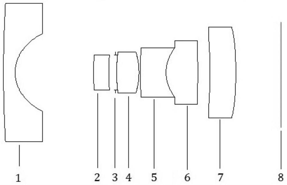
[0027] The electronic hysteroscopic imaging lens of the present invention selects a five-element "inverse telephoto" lens group with negative focal power in the front and positive focal power in the rear as the initial structure, which can take into account both a large field of view and a large relative aperture. With a longer working distance, compared with the double Gaussian structure, the illumination of the image plane is relatively uniform.

[0028] like figure 1 As shown, the electronic hysteroscopic imaging lens of the present invention includes a first lens 1, a second lens 2, a third lens 4, a fourth lens 5, a fifth lens 6 and a sixth lens arranged in sequence from the object to the image 7. A diaphragm 3 is placed between the second lens 2 and the third lens 4 , the fourth lens 5 and the fifth lens 6 form a doublet lens, and the i...


PUM


Abstract

The invention discloses a dual-purpose electronic hysteroscope imaging lens for distending uterus by gas and liquid, which comprises a first lens, a second lens, a third lens, a fourth lens, a fifth lens and a sixth lens which are sequentially arranged from object to image, a diaphragm is arranged between the second lens and the third lens, the fourth lens and the fifth lens form a doublet lens, and an image plane is arranged outside the sixth lens. The lens is small in aperture, can obviously relieve pain of a patient, and is suitable for early diagnosis of uterine cancer; the structure is simple, operation is convenient, and the multi-scene application requirement can be met only by adjusting the intervals of the lens sets; 90-degree view field non-blind area, large-depth-of-field and high-resolution imaging can be achieved, the detection rate and the definite diagnosis rate of early diseases, especially tumors, are improved, unnecessary operation of doctors on instruments in the body is reduced, and operation risks are reduced.

Description

technical field [0001] The invention relates to the field of optical technology, in particular to an electronic hysteroscopic imaging lens for both gas and liquid uterine expansion, which is used for uterine cavity imaging during gynecological minimally invasive surgery and hysteroscopic anesthesia-free examination. Background technique [0002] With the gradual improvement of living standards, people pay more and more attention to their own health. Among the malignant tumors of women in the world, the incidence of cervical cancer ranks second. It takes more than ten years for cervical cells to mutate into cancer cells. If the disease can be detected and treated in time at the early stage, the cure rate can reach 100%. At present, in clinical medicine, the diagnostic methods of uterine diseases mainly include ultrasonic technology and medical endoscope. Traditional ultrasonic technology can only observe the section of organ fault structure, which has the disadvantages of in...

Claims


Application Information

Patent Timeline
IPC IPC(8): G02B23/24G02B13/00G02B13/06G02B13/18A61B1/303A61B1/00
CPCG02B23/243G02B13/0045G02B13/006G02B13/06G02B13/18A61B1/303A61B1/00177
Inventor 武旭华刘国栋
Owner NANJING UNIV OF INFORMATION SCI & TECH
Learn More