
Bone marrow smear microscopic image analysis method and device, electronic equipment and storage medium

A microscopic image and bone marrow smear technology, which is applied in the field of image processing, can solve problems such as false detection and missed detection, and achieve the effect of improving accuracy

Pending Publication Date: 2021-02-26
SHANGHAI UNITED IMAGING INTELLIGENT MEDICAL TECH CO LTD
View PDF0 Cites 0 Cited by
  • Summary
  • Abstract
  • Description
  • Claims
  • Application Information

AI Technical Summary

Problems solved by technology

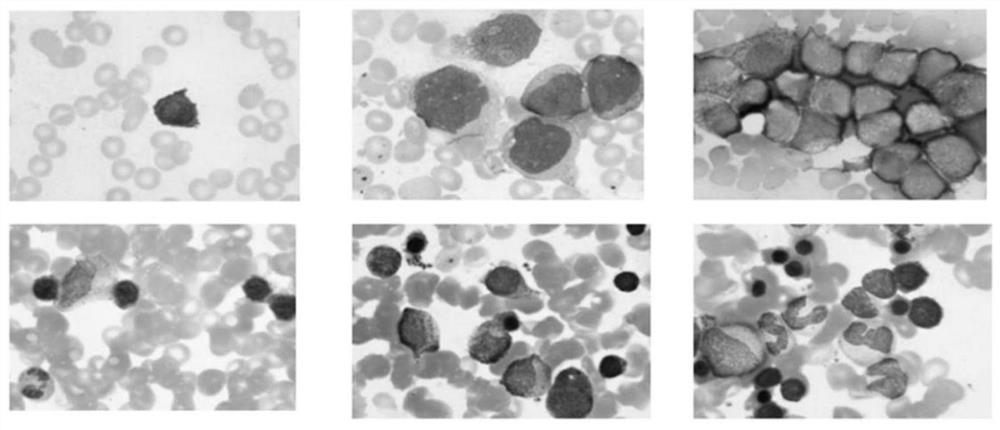
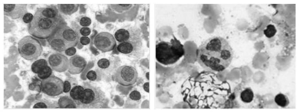
[0005] The technical problem to be solved by the present invention is to overcome the fact that in the prior art, when detecting bone marrow cells, it is easy to treat bone marrow cells with serious adhesion and occlusion as one bone marrow cell, resulting in missed detection, and to treat special cells with multiple nucleoli as two cells. However, the defects that lead to false detection provide a method, device, electronic equipment and storage medium for analyzing microscopic images of bone marrow smears.

Method used


Image

  • Bone marrow smear microscopic image analysis method and device, electronic equipment and storage medium
  • Bone marrow smear microscopic image analysis method and device, electronic equipment and storage medium
  • Bone marrow smear microscopic image analysis method and device, electronic equipment and storage medium

Examples


Embodiment 1

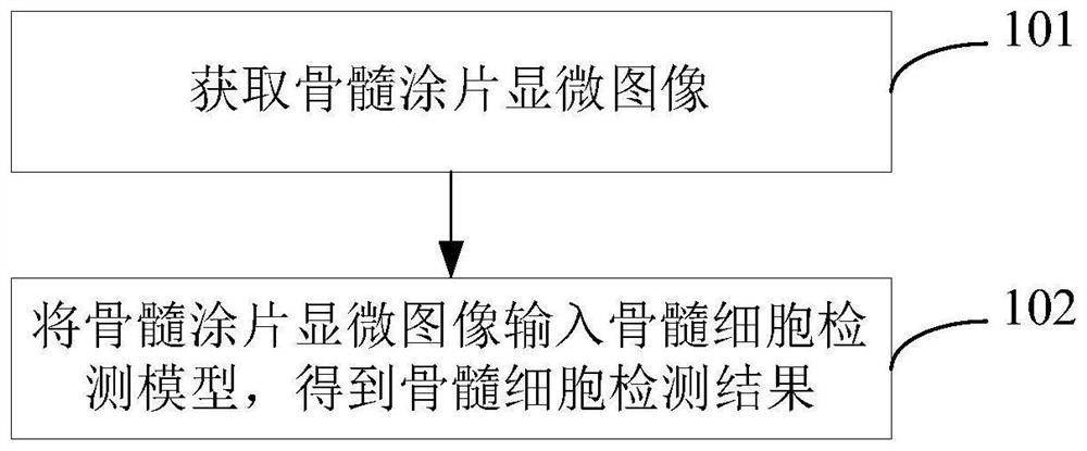
[0059] This embodiment provides a method for analyzing a microscopic image of a bone marrow smear. Among them, the microscopic image of bone marrow smear refers to the image of bone marrow smear under the microscopic field of view, where the bone marrow smear can be the original, unprocessed bone marrow smear, or after Wright's staining or other treatments. Bone marrow smear, which may contain bone marrow cells. Such as image 3 As shown, it includes the following steps:

[0060] Step 101: Obtain a microscopic image of the bone marrow smear.

[0061] Step 102: Input the microscopic image of the bone marrow smear into the bone marrow cell detection model to obtain the bone marrow cell detection result. Among them, the bone marrow cell detection model is used to predict the position distribution map of bone marrow cells in the microscopic image of bone marrow smear, and the position distribution map is fused with the original feature map extracted from the microscopic image o...

Embodiment 2

[0079] This embodiment is a further improvement on Embodiment 1. The method of this embodiment can further realize the classification of bone marrow cells. The classification may include primary classification according to four types of lineage cells and secondary classification according to cells in different growth stages.

[0080] Bone marrow cells proliferate under the action of regulatory factors, which is an important basis for growth and development. The four cell lineages are granulocytes, nucleated erythrocytes, lymphocytes, and monocytes. According to different growth stages, granulocytes are divided into myeloblasts, promyelocytes, neutrophils, eosinophils, and basophils; nucleated erythrocytes are divided into primitive erythrocytes, promyelocytes, and mesenchyme , late immature red blood cells; lymphocytes are divided into primitive lymphocytes, immature lymphocytes, mature lymphocytes and plasma cells; monocytes are divided into primitive monocytes, immature mo...

Embodiment 3

[0096] This embodiment provides a microscopic image analysis device for a bone marrow smear. Among them, the microscopic image of bone marrow smear refers to the image of bone marrow smear under the microscopic field of view, where the bone marrow smear can be the original, unprocessed bone marrow smear, or after Wright's staining or other treatments. Bone marrow smear, which may contain bone marrow cells. Figure 8 A schematic block diagram of the device is shown, which includes: an image acquisition module 301 and a cell detection module 302 .

[0097] The image acquisition module 301 is used for acquiring a microscopic image of a bone marrow smear.

[0098] The cell detection module 302 is used to input the microscopic image of the bone marrow smear into the bone marrow cell detection model to obtain the bone marrow cell detection result. Among them, the bone marrow cell detection model is used to predict the position distribution map of bone marrow cells in the microscop...


PUM


Abstract

The invention discloses a bone marrow smear microscopic image analysis method and device, electronic equipment and a storage medium. The method comprises the following steps: acquiring a bone marrow smear microscopic image; inputting the bone marrow smear microscopic image into a bone marrow cell detection model to obtain a bone marrow cell detection result; wherein the bone marrow cell detectionmodel is used for predicting a position distribution diagram of bone marrow cells in the bone marrow smear microscopic image, fusing the position distribution diagram with an original feature map extracted from the bone marrow smear microscopic image to obtain fusion features, and outputting a bone marrow cell detection result according to the fusion features. The method does not depend on subjective judgment of medical personnel and has the advantages of being objective and efficient, prediction of the position distribution diagram is introduced into the bone marrow cell detection model, extracted semantic features are enriched, missing detection and wrong detection are not likely to happen, and the bone marrow cell detection accuracy is improved. Comprehensive analysis such as positioning, recognition, classification and statistics of bone marrow cells is further realized.

Description

technical field [0001] The invention belongs to the field of image processing, and in particular relates to a bone marrow smear microscopic image analysis method, device, electronic equipment and storage medium. Background technique [0002] Morphological examination of bone marrow cells is the earliest application of a method for the classification and diagnosis of blood tumors, and it is also one of the most basic and important methods at present. However, at present, this microscopic examination method still adopts the traditional way of manual observation, classification and counting. This method is inefficient, labor-intensive, has high requirements for medical experts, and the recognition results depend on human subjective judgment. Therefore, with the help of image processing and artificial intelligence technology, it is of great significance to improve the efficiency and level of clinical diagnosis of diseases by analyzing the microscopic images of bone marrow smear...

Claims


Application Information

Patent Timeline
IPC IPC(8): G06T7/00G06T7/90G06T5/00G06K9/62G06N3/04G06N3/08
CPCG06T7/0012G06T7/90G06N3/08G06T2207/10061G06T2207/30242G06N3/045G06F18/2414G06F18/253G06T5/70
Inventor 刘慧敏沈宏
Owner SHANGHAI UNITED IMAGING INTELLIGENT MEDICAL TECH CO LTD
Learn More