Eureka AIR delivers breakthrough ideas for toughest innovation challenges, trusted by R&D personnel around the world.

Automatic focus searching method and system for pathological images under microscope

A pathological image, a technique under the microscope

Active Publication Date: 2021-02-12
杭州医派智能科技有限公司
View PDF18 Cites 2 Cited by
  • Summary
  • Abstract
  • Description
  • Claims
  • Application Information

AI Technical Summary

Problems solved by technology

[0004] Existing digital pathological slide scanners have a slice scanning part and a slice replacement part, but the existing digital pathological slide scanners basically use a computer to control the work of the scanner In this way, this kind of scanning usually does not have a fixed-focus process. In this case, to obtain a sufficiently cleaned pathological image becomes the technical bottleneck of the digital slicer. At present, the fixed-focus process still needs to be manually or pre-set. , manual focus will undoubtedly reduce work efficiency, and the pre-set method has very high requirements for the initial prevention position of the pathological image. If the placement position of the pathological image is inaccurate or even slightly wrong, it will not be able to focus accurately

Method used


Image

  • Automatic focus searching method and system for pathological images under microscope
  • Automatic focus searching method and system for pathological images under microscope
  • Automatic focus searching method and system for pathological images under microscope

Examples


Embodiment 1

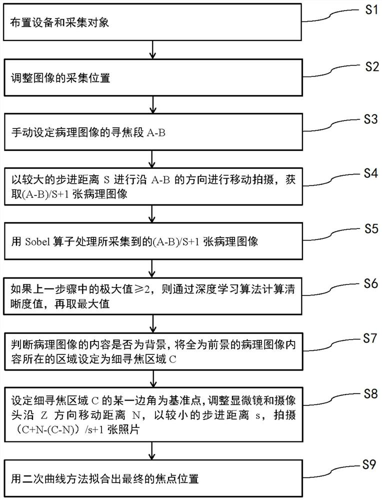
[0039] Example 1, with reference to the attached figure 1 .

[0040] In one embodiment, as figure 1As shown, the present invention provides an automatic focusing method for pathological images under a microscope, the method can be used for automatic focusing in the process of collecting pathological images under a microscope by a digital slicer, and specifically includes the following steps:

[0041] S1, arranging equipment and collection objects: place the microscope and camera facing the collection area, place the slide containing the pathological slices in the collection area, and check whether the equipment is functioning normally;

[0042] The collection diagram after setting is as follows Figure 5 where the coordinate system corresponds to two mutually perpendicular directions in the plane where the acquisition area is located according to the X axis and the Y axis, and the Z axis corresponds to where the acquisition device (microscope and camera) is located and is mo...

Embodiment 2

[0058] Example 2, with reference to the attached figure 2 .

[0059] In one embodiment, as figure 2 As shown, the present invention provides an automatic focusing system for pathological images under a microscope. The automatic focusing system includes a central processing unit 100, an acquisition module 200 and an adjustment module 300. The acquisition module 200 is as described above. It can include a microscope and a camera. The present invention does not need to improve the selection and installation of the microscope and the camera, because the specific installation method and hardware selection can adopt the solutions in the prior art. There is no need to make improvements in the combined use of , and the method in the digital film reading technology in the prior art can be adopted, so the technical content of this piece is not described in detail in the present invention.

[0060] The acquisition module 200 is used to collect pathological images of a designated area...

Embodiment 3

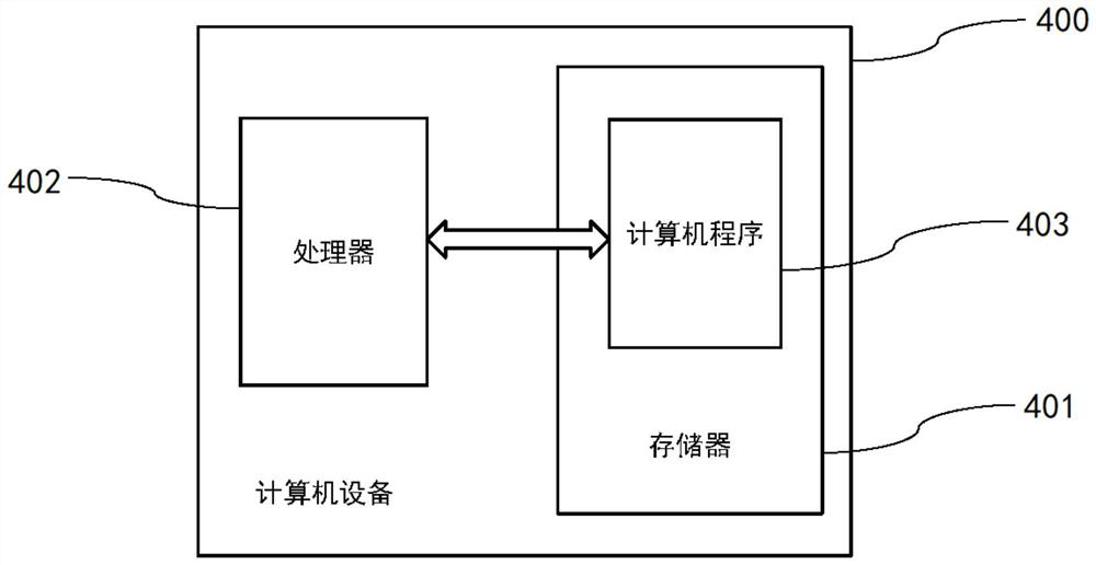
[0061] Example 3, with reference to the attached image 3 .

[0062] In this embodiment, a computer device 400 is provided, the computer device 400 includes a memory 401, a processor 402, and a computer program 403 stored in the memory 401 and executable on the processor 402, and the processor 402 executes the computer program At 403 , the steps in the automatic focusing method provided in the above Embodiment 1 can be implemented.

[0063] In addition, the relationship between the processor 402 in this embodiment and the central processing unit in Embodiment 1 and the central processing unit 200 in Embodiment 2 may be that the processor 402 includes the central processing unit 200, or the two have common Execution or functional modules, or both, are two mutually independent processors that can communicate with each other. Actually, the processor 402 in this embodiment can also execute the functions of the central processing unit in the first embodiment and the central proce...


PUM


Abstract

The invention provides an automatic focus searching method for pathological images under a microscope. The automatic focus searching method comprises the steps: arranging equipment and an acquisitionobject; adjusting the acquisition position of an image; roughly positioning a pathological image; and carrying out fine focus searching on the pathological image to obtain a final focus position. According to the automatic focus searching method, a preliminary positioning process and a fine positioning process exist, the step-by-step cleaning process of focus searching is met, meanwhile, operationis conducted through a definition algorithm in the preliminary positioning process and the fine positioning process, and the accuracy of automatic focus searching is improved under the condition thatthe processing speed is not affected.

Description

technical field [0001] The invention relates to the technical field of detection and identification, and more particularly, to a method for automatic focusing of pathological images under a microscope. Background technique [0002] Pathological examination is a pathological morphological method used to examine pathological changes in body organs, tissues or cells. In order to explore the disease process of organs, tissues or cells, a certain pathological morphological examination method can be used to examine their lesions, to explore the causes, pathogenesis, and development of lesions, and finally to make a pathological diagnosis. diagnosis. The pathological examination method is to first observe the pathological changes of the gross specimen, then cut out a certain size of the diseased tissue, make pathological sections with histopathological methods, and further examine the lesions with a microscope. [0003] The digital slide system can quickly scan the entire slide w...

Claims


Application Information

Patent Timeline
Patent Type & Authority Applications(China)
IPC IPC(8): G02B21/24
CPCG02B21/241
Inventor 汪太平张敏飞
Owner 杭州医派智能科技有限公司
Why Eureka
Eureka Blog
Learn More