
Imaging physician anti-ray protection device

A protection device and radiation-proof technology, applied in the medical field, can solve the problems of affecting patient communication, inability to adjust the height of radiation-proof glass with different doctors, and hazards of radioactive substances to doctors, etc., and achieve the effect of overall stability.

Pending Publication Date: 2021-02-05
重庆见芒信息技术咨询服务有限公司
View PDF0 Cites 1 Cited by
  • Summary
  • Abstract
  • Description
  • Claims
  • Application Information

AI Technical Summary

Problems solved by technology

[0003] At present, the imaging department of the hospital often uses radiation to check the diseases of the internal organs of certain patients. The relevant personnel must be exposed to radiation for a long time, especially when the patient is undergoing PET nuclide imaging examination, radioactive substances will be injected. After the examination is completed, the radioactive substances will remain in the patient's body for a period of time. When the patient communicates with the doctor, the radioactive substances on the patient's body will cause certain harm to the doctor.
[0004] The anti-ray glass of the existing anti-ray protection table is usually fixed, and the height of the anti-ray glass cannot be adjusted according to the height of different doctors. If the anti-ray glass is too high, it will affect the communication with patients; The range of radiation becomes smaller, which makes doctors affected by radiation and cannot meet the needs of users

Method used


Image

  • Imaging physician anti-ray protection device
  • Imaging physician anti-ray protection device
  • Imaging physician anti-ray protection device

Examples


Embodiment Construction

[0033] In order to enable those skilled in the art to better understand the present invention, the technical solution of the present invention will be further described below in conjunction with the accompanying drawings and embodiments.

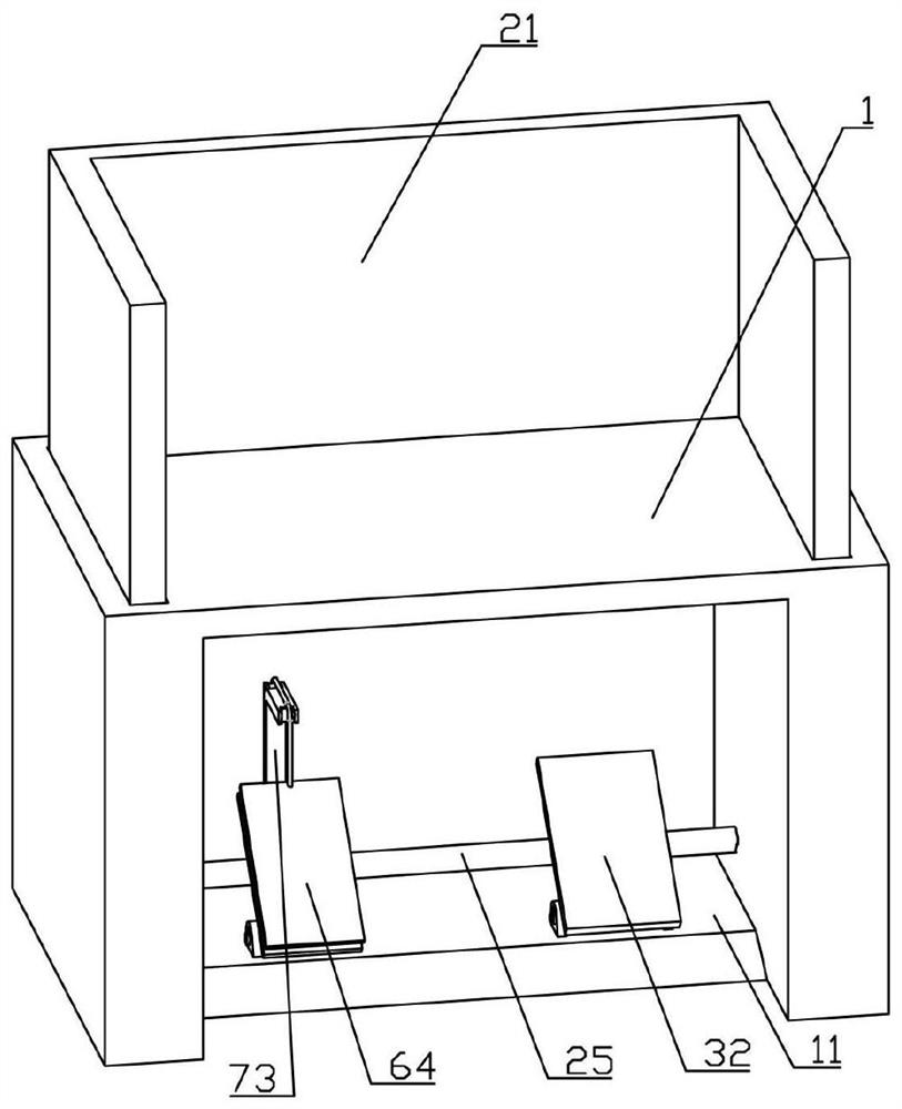
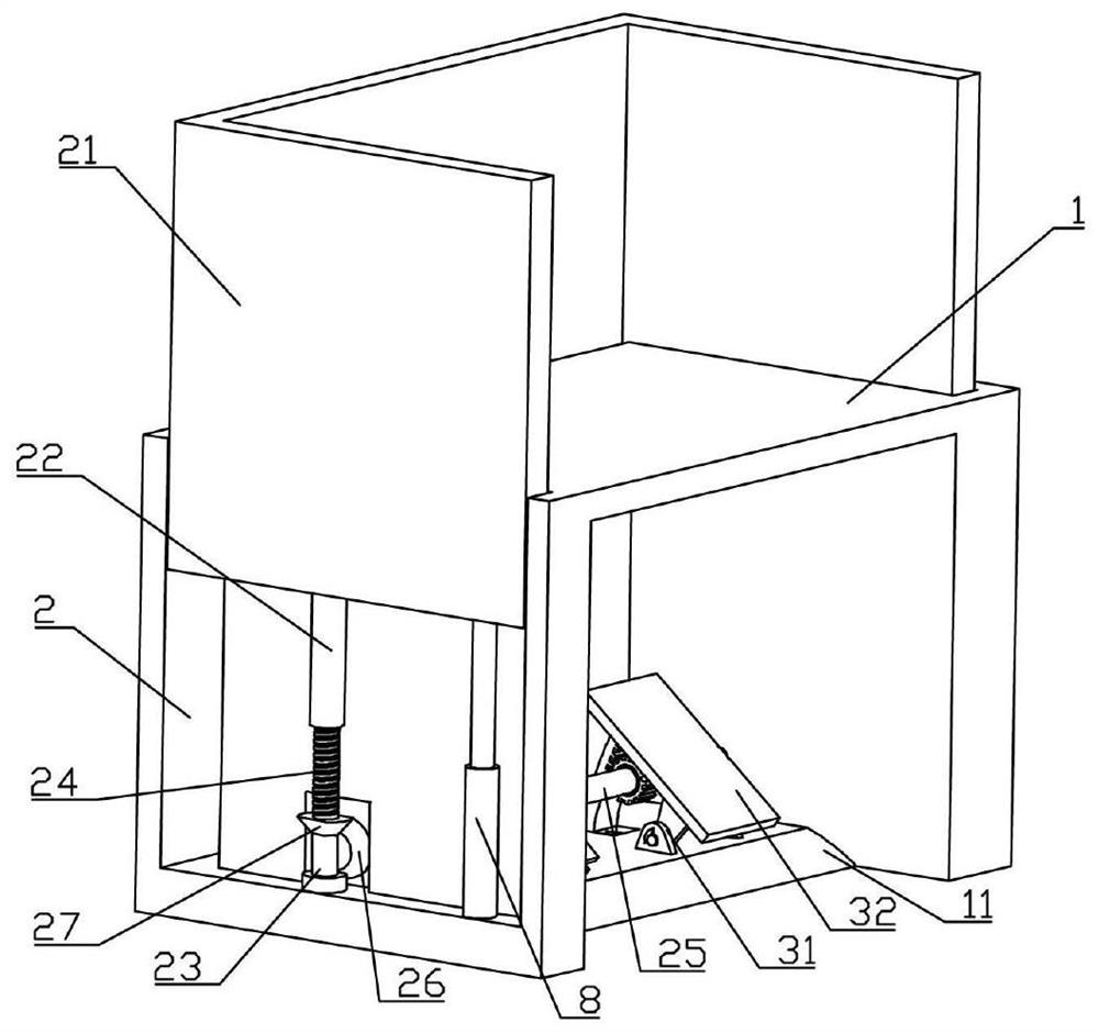
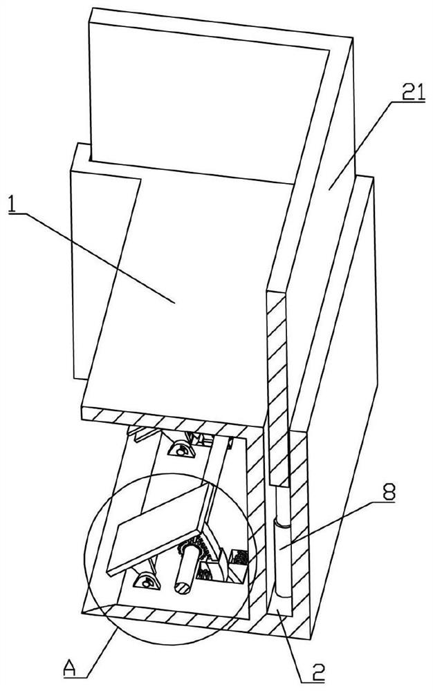
[0034] Such as Figure 1-8 As shown, a radiation protection device for radiologists of the present invention includes a protection table 1, a mounting plate 11 is fixedly connected to the bottom of the protection table 1, and a U-shaped groove 2 is provided on the protection table 1, and the U-shaped groove 2 is slidably connected U-shaped anti-ray glass 21 is arranged, the left and right sides of the bottom of U-shaped anti-ray glass 21 are fixedly connected with threaded sleeve 22, the bottom of U-shaped groove 2 is rotatably connected with support seat 23, and the support seat 23 is fixedly connected with threaded sleeve 22 threads The connected threaded rod 24 is connected to the rotating shaft 25 in the middle of the protection table 1,...


PUM


Abstract

The invention belongs to the technical field of medical treatment, and particularly relates to an imaging physician anti-ray protection device which comprises a protection table. A U-shaped groove isformed in the protection table, U-shaped anti-ray glass is arranged in the U-shaped groove, a threaded sleeve is arranged on the U-shaped anti-ray glass, a threaded rod is arranged on the threaded sleeve, a rotating shaft is arranged on the protection table, a first gear is arranged on the rotating shaft, a first elastic hinge seat is arranged on a mounting plate, the first elastic hinge seat is provided with a first pedal, the rotating shaft is provided with a second gear, the mounting plate is provided with a second elastic hinge seat, the second elastic hinge seat is fixedly connected witha second pedal, the second pedal is provided with an elastic sheet, and the elastic sheet is provided with a pressing plate; A physician can adjust the height of the U-shaped anti-ray glass only by stepping on the first pedal or the second pedal, so that the height of the anti-ray glass is adjusted according to the heights of different physicians, the physicians are effectively prevented from being affected by rays, convenience is brought to work of the physician, the use requirements of users are met, operation is easy, and practicability is high.

Description

technical field [0001] The invention belongs to the field of medical technology, and in particular relates to a radiation protection device for radiologists. Background technique [0002] Imaging not only expands the scope of human body examination and improves the level of diagnosis, but also can treat certain diseases. In this way, the work content of this discipline has been greatly expanded, and it has become an important pillar in medical work. Shortly after Roentgen discovered X-rays in 1895, X-rays have been used in medicine to examine the human body and diagnose diseases, forming a new discipline of radiodiagnostics and laying the foundation for medical imaging. So far, diagnostic radiology is still the main content of medical imaging, and it is widely used. [0003] At present, the imaging department of the hospital often uses radiation to check the internal organs of some patients. The relevant personnel must be exposed to radiation for a long time, especially wh...

Claims


Application Information

Patent Timeline
Patent Type & Authority Applications(China)
IPC IPC(8): G21F3/00A61B6/10
CPCG21F3/00A61B6/10
Inventor 皮历魏祯奇王浩南李冬谢旭王潇刘余刘朝燕
Owner 重庆见芒信息技术咨询服务有限公司
Learn More