Transabdominal preperitoneal hernia repair operation training model and its production method and preservation method
A production method and training model technology, applied in the medical field, can solve problems such as high investment, large differences between animals and humans, and inability to meet human surgery, and achieve the effects of low cost, convenient material acquisition, and simple training environment requirements
- Summary
- Abstract
- Description
- Claims
- Application Information
AI Technical Summary
Problems solved by technology
Method used
Image
Examples
Embodiment 1
[0092] A transabdominal preperitoneal hernia repair operation training model, the production method comprising the following steps:
[0093]1. Take the stomach tissue of the pig along the lesser curvature to remove the wrinkled and unsmooth gastric body of the lesser curvature of the stomach;
[0094] 2. Cut the gastric fundus and gastric body of the gastric tissue to obtain the gastric fundus and gastric body;
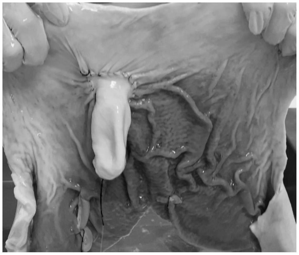
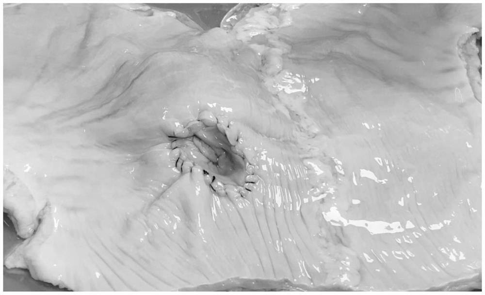
[0095] 3. The schematic diagram of gastric fundus is as follows: image 3 As shown, the mucosal layer and the seromuscular layer at the edge of the gastric fundus were separated by 0.5 cm; then the long axis AB of the gastric fundus was used as the folding line to fold in half, and the edge of the butt-joined mucosal layer was sutured from the A end to the B end, and the diameter of the B end was retained. 2.5cm opening; then two chicken intestines with a diameter of 0.8cm and a length of 12cm were placed between the separate mucosal layer and the seromuscular layer,...
Embodiment 2
[0101] A transabdominal preperitoneal hernia repair operation training model, the production method comprising the following steps:
[0102] 1. Take the stomach tissue of the sheep along the lesser curvature to remove the wrinkled and unsmooth gastric body of the lesser curvature of the stomach;
[0103] 2. Cut the gastric fundus and gastric body of the gastric tissue to obtain the gastric fundus and gastric body;
[0104] 3. Separate the mucosal layer and the seromuscular layer at the edge of the gastric fundus by 1 cm; then fold in half with the long axis AB of the gastric fundus as the folding line, and fix the edge of the butt-joined mucosal layer from the A end to the B end with a fixed pin and keep the diameter at the B end Then put two duck intestines with a diameter of 1cm and a length of 15cm between the separated mucosal layer and seromuscular layer, and one end of the duck intestine was sutured and fixed at the A end, and the inside of the duck intestine was filled ...
Embodiment 3
[0110] A transabdominal preperitoneal hernia repair operation training model, the production method comprising the following steps:
[0111] 1. Take the gastric tissue of the cow along the lesser curvature to reduce the wrinkled and uneven gastric body of the lesser curvature of the stomach;
[0112] 2. Cut the gastric fundus and gastric body of the gastric tissue to obtain the gastric fundus and gastric body;
[0113] 3. Separate the mucosal layer and seromuscular layer at the edge of the gastric fundus by 1.5 cm; then fold in half with the long axis AB of the gastric fundus as the folding line, suture the edge of the butt-joined mucosal layer from end A to end B, and keep the diameter of end B at 3.5 cm opening; then two chicken intestines with a diameter of 1.2 cm and a length of 18 cm were placed between the separated mucosal layer and seromuscular layer, and one end of the chicken intestine was fixed on the A end, and the inside of the chicken intestine was filled with wa...
PUM
 Abstract
Description
Claims
Application Information
 - R&D
- Intellectual Property
- Life Sciences
- Materials
- Tech Scout
- Unparalleled Data Quality
- Higher Quality Content
- 60% Fewer Hallucinations
Browse by: Latest US Patents, China's latest patents, Technical Efficacy Thesaurus, Application Domain, Technology Topic, Popular Technical Reports.
© 2025 PatSnap. All rights reserved.Legal|Privacy policy|Modern Slavery Act Transparency Statement|Sitemap|About US| Contact US: help@patsnap.com


