Cartilago articularis quantitative imaging analysis method based on T2 mapping and T1 rho

A technology of articular cartilage and analysis methods, applied in image analysis, image enhancement, image data processing, etc.

Pending Publication Date: 2020-12-25
QINGDAO MUNICIPAL HOSPITAL
View PDF2 Cites 0 Cited by
  • Summary
  • Abstract
  • Description
  • Claims
  • Application Information

AI Technical Summary

Problems solved by technology

At present, many researchers at home and abroad are also studying the influencing factors of T1ρ value, but there are many controversies

Method used


Image

  • Cartilago articularis quantitative imaging analysis method based on T2 mapping and T1 rho
  • Cartilago articularis quantitative imaging analysis method based on T2 mapping and T1 rho

Examples


Embodiment Construction

[0020] A method for quantitative imaging and analysis of articular cartilage based on T2 mapping and T1ρ of the present invention will be described in detail below.

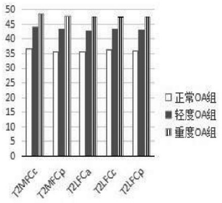
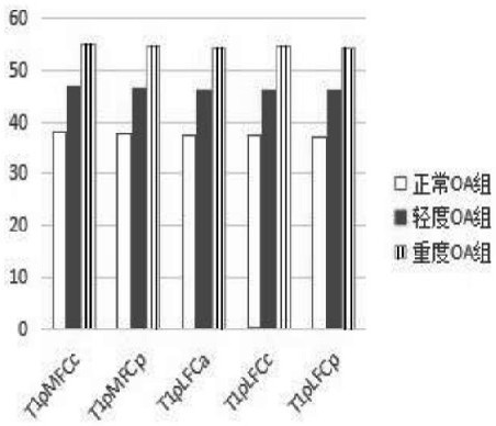
[0021] Such as figure 1 and figure 2 As shown, a quantitative imaging analysis method based on T 2mapping and T1ρ cartilage. The steps are as follows: 1. MRI scanning: use GE 3.0T (MR750) magnetic resonance imager and knee joint special coil (HD TRknee) to complete the examination. The patient is placed in the supine position, the left or right knee is scanned, the foot is advanced, the knee joint is extended, the lower edge of the patella is the scanning center, and the scanning range is from the level of the upper edge of the patella to the tibial plateau. Since exercise or weight-bearing may cause temporary changes in articular cartilage, patients and normal volunteers should ensure that they do not engage in strenuous activities and sit quietly for at least 30 minutes before being examined;

[0022] 2. Di...


PUM


Abstract

The invention discloses a cartilago articularis quantitative imaging analysis method based on T2 mapping and T1 rho. The cartilago articularis quantitative imaging analysis method comprises the following steps: 1, carrying out MRI scanning; 2, dividing a lower end area of the femur; 3, conducting image evaluation; 4, measuring data; 5, carrying out statistical analysis, wherein the scanning sequence and parameters of the MRI scanning comprise conventional scanning and non-conventional scanning. According to the articular cartilage quantitative imaging analysis method based on T2 mapping and T1rho, knee osteoarthritis is a multi-factor disease, many patients suffer from the disease, and the social burden is heavy. A new strategy for treating knee osteoarthritis is early discovery and minimally invasive treatment, T2 and T1 rho values can be measured through magnetic resonance T2 mapping and T1 rho imaging technologies, change of an articular cartilage biochemical structure is reflectedfrom the molecular level before morphological change of articular cartilage, high sensitivity and specificity are achieved, and early diagnosis and treatment can be conducted on early OA.

Description

technical field [0001] The invention relates to a method for quantitative imaging analysis of articular cartilage, in particular to a method for quantitative imaging analysis of articular cartilage based on T2 mapping and T1ρ. Background technique [0002] Quantitative T1ρ imaging is a new MRI imaging technique that has emerged in recent years. T1ρ uses a spin-lock pulse sequence to magnetize before the standard fast spin echo, sets the spin-lock time, and reconstructs the T1ρ image, which is represented by a color-scale or gray-scale image, and different ROIs can be measured on the image The T1ρ value is used to evaluate the spin relaxation value of the tissue in the pulsed magnetic field, analyze the changes of the tissue components in the cartilage, and make a diagnosis of early OA. [0003] T1ρ imaging is an examination method for probing the interaction between macromolecular species and water molecules during slow motion. Early cartilage degeneration in OA is due to ...

Claims


Application Information

Patent Timeline
IPC IPC(8): G06T7/00A61B5/055
CPCG06T7/0012A61B5/055G06T2207/10088G06T2207/30008
Inventor 张炳陈鑫王国华
Owner QINGDAO MUNICIPAL HOSPITAL
Why Eureka
Try Eureka