
Magnetic anchoring pulmonary nodule positioning device for thoracoscopic surgery

A positioning device and thoracoscopic technology, applied in the field of medical devices, can solve the problems of increased complications, high price, and lack of intuitiveness, and achieve the effects of reducing discomfort and complications, preventing pathological judgment, and avoiding direct contact

Active Publication Date: 2020-10-27
崇好科技有限公司
View PDF8 Cites 1 Cited by
  • Summary
  • Abstract
  • Description
  • Claims
  • Application Information

AI Technical Summary

Problems solved by technology

However, for pulmonary nodules with a subpleural depth of more than 5 mm and a diameter of less than 10 mm, especially pure ground-glass nodules, conventional methods such as vision, finger palpation, or surgical instrument sliding cannot be accurately located during thoracoscopic surgery, which may lead to prolonged operation time. Prolonged, excessive resection of normal lung tissue, etc., which in turn affect the curative effect of surgery, increase postoperative complications, are not conducive to the recovery of patients, and increase the possibility of conversion to thoracotomy, which deviates from the fundamental starting point of thoracoscopic surgery
In order to optimize the application of thoracoscopy in pulmonary nodule resection, a series of pulmonary nodule localization techniques have been clinically explored in recent years, such as puncture localization technology, image-mediated localization technology, electromagnetic navigation bronchoscopy localization, etc. Displacement or falling off, unintuitive, expensive defect

Method used


Image

  • Magnetic anchoring pulmonary nodule positioning device for thoracoscopic surgery
  • Magnetic anchoring pulmonary nodule positioning device for thoracoscopic surgery
  • Magnetic anchoring pulmonary nodule positioning device for thoracoscopic surgery

Examples


Embodiment Construction

[0023] The implementation of the present invention will be described in detail below in conjunction with the drawings and examples.

[0024] The present invention is a magnetically anchored pulmonary nodule positioning device for thoracoscopic surgery, comprising:

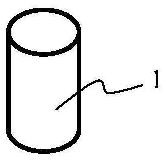
[0025] Two target magnets 1 are used to clamp the target nodule 7 on both sides of the target nodule 7, refer to figure 1 , in this embodiment, the two target magnets 1 have the same specifications, are columnar magnets, and the magnetic poles are radial in direction, and attract each other in the radial direction after clamping the target nodule 7. The material of the target magnet 1 can be coated NdFeB.

[0026] Two coaxial trocar needles 2, ref. figure 2 , in this embodiment, the two coaxial puncture needles 2 are hollow cylindrical structures with the same specification, and their inner diameter is slightly larger than the outer diameter of the target magnet 1, for the target magnet 1 to be fed in the axial d...


PUM


Abstract

The invention provides a magnetic anchoring pulmonary nodule positioning device for thoracoscopic surgery. The device comprises: two target magnets, which are used for clamping a target nodule on twosides of the target nodule; two coaxial puncture needles, which are of hollow structures, and the hollow sizes of the two coaxial puncture needles are larger than the external sizes of the two targetmagnets respectively so as allow the two target magnets to penetrate through the two coaxial puncture needles respectively; a positioning plate, which is provided with a plurality of holes, wherein two coaxial puncture needles penetrate into two of the holes to realize preliminary positioning of a target nodule; and an anchoring magnet, which is used for being attracted with the target magnets onthe lung surface through magnetic force, and a positioning range is determined according to the magnetic force. According to the invention, nodule lesions are positioned through magnetic attraction force. The device is suitable for pulmonary lobe excision under a thoracoscope. Meanwhile, the probability that a positioning marker falls off, shifts and moves is lowered, stimulation of a foreign matter to the pleura can be reduced, corresponding discomfort and complications are relieved, the marker is prevented from making direct contact with the nodule, pathological judgment in an operation is prevented from being affected, and the requirements of more patients for high-precision thoracoscopic surgery are met.

Description

technical field [0001] The invention belongs to the technical field of medical devices, in particular to a magnetically anchored pulmonary nodule positioning device for thoracoscopic surgery. Background technique [0002] As the main technical method for the diagnosis and treatment of pulmonary nodules, thoracoscopic technology has the advantages of less trauma, faster recovery, and fewer surgical complications, and is widely used in clinical practice. However, for pulmonary nodules with a subpleural depth of more than 5 mm and a diameter of less than 10 mm, especially pure ground-glass nodules, conventional methods such as vision, finger palpation, or surgical instrument sliding cannot be accurately located during thoracoscopic surgery, which may lead to prolonged operation time. Prolonged, excessive resection of normal lung tissue, etc., which in turn affect the curative effect of surgery, increase postoperative complications, are not conducive to the recovery of patients,...

Claims


Application Information

Patent Timeline
IPC IPC(8): A61B90/00
CPCA61B90/39A61B2090/3908A61B2090/3954
Inventor 崔晓海张靖吕毅慕凡刘大鹏付军科张云锋
Owner 崇好科技有限公司
Learn More