Immunofluorescence kit for detecting PD-L1 expression of peripheral blood circulating tumor cells of kidney cancer patient and detection method

A PD-L1 and peripheral blood circulation technology, applied in tumor/cancer cells, cell dissociation methods, animal cells, etc., can solve the problem that tissue samples cannot evaluate the real-time dynamic status of PD-L1 in patients, false negatives, false positives in detection methods, etc. problems, to avoid edge effects, reduce cell loss, and achieve good stability

Inactive Publication Date: 2020-08-11
SHANDONG FIRST MEDICAL UNIV & SHANDONG ACADEMY OF MEDICAL SCI
View PDF0 Cites 3 Cited by
  • Summary
  • Abstract
  • Description
  • Claims
  • Application Information

AI Technical Summary

Problems solved by technology

[0008] Aiming at the disadvantages of detecting patients with advanced tumors or recurrent renal cancer in real-time or repeated punctures to obtain tissue samples in the prior art, and thus not being able to evaluate the real-time dynamic status of PD-L1 in patients, and the existing detection methods are prone to false positives and false negatives, this paper The invention provides an immunofluorescence kit and detection method for the expression of PD-L1 in the peripheral blood of patients with renal cancer, using a membrane filtration device to separate and obtain circulating tumor cells (CTCs) in the peripheral blood of patients with advanced renal cancer, and further using immunofluorescence Technology detection of PD-L1 expression on CTC

Method used


Image

  • Immunofluorescence kit for detecting PD-L1 expression of peripheral blood circulating tumor cells of kidney cancer patient and detection method
  • Immunofluorescence kit for detecting PD-L1 expression of peripheral blood circulating tumor cells of kidney cancer patient and detection method
  • Immunofluorescence kit for detecting PD-L1 expression of peripheral blood circulating tumor cells of kidney cancer patient and detection method

Examples


Embodiment 1

[0050] 1. Use a membrane filtration device to separate and obtain CTCs in the peripheral blood of patients with advanced or recurrent renal cancer whose tissue samples cannot be obtained, and determine whether CTCs exist:

[0051] Collect 5ml of fasting blood from the median cubital vein for 8-12 hours, dilute the peripheral blood with 45ml diluent (ingredients: 1mmol / L EDTA+0.1%BSA+0.1%trehalose+0.2% lauryl polyoxyethylene ether), and then Add 3ml of 4% paraformaldehyde to fix the diluted blood sample for 10 minutes;

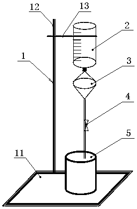
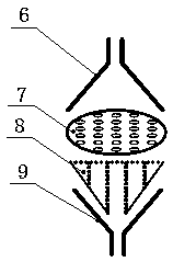
[0052] At fixed intervals, assemble the membrane filtration device: as attached figure 1 , figure 2 , image 3 As shown, the filtering device is composed of a filter 3, a filter membrane 7, a blood sample container 2, a waste liquid tank 5, and an iron stand 1;

[0053] Wet the filter 3 with 10ml of PBS, and then add the fixed peripheral blood sample into the blood sample container 2 of the membrane filtration device, so that it is naturally filtered by gra...


PUM

PropertyMeasurementUnit
Diameteraaaaaaaaaa
Diameteraaaaaaaaaa

Abstract

The invention belongs to the technical field of molecular biology, and particularly relates to an immunofluorescence kit for detecting PD-L1 expression of peripheral blood circulating tumor cells of akidney cancer patient and a detection method. The kit comprises 45mL of a diluent, 1mL of a destaining solution, 0.5mL of staining solution A, 1mL of a staining solution B, 200[mu]l of methanol, 200[mu]l of 2% PFA, 100[mu]l of 10% goat serum, 100[mu]l of a primary antibody suspension composed of mouse anti-CK, rat anti-CD45 and rabbit anti-PD-L1, 100[mu]l of a secondary antibody suspension composed of a fluorescently labeled goat anti-mouse, a fluorescently labeled goat anti-rat and a fluorescently labeled goat anti-rabbit, and a DAPI mounting medium. According to the detection method provided by the invention, the PD-L1 expression condition of a patient with advanced or recurrent renal cancer can be detected without acquiring a tissue specimen through needle biopsy. The technology belongs to minimally invasive and can realize real-time detection.

Description

technical field [0001] The invention belongs to the technical field of molecular biology, and in particular relates to an immunofluorescence kit and a detection method for detecting the expression of PD-L1 in peripheral blood circulating tumor cells of kidney cancer patients. Background technique [0002] Renal cell carcinoma (RCC) is one of the most common malignant tumors of the urinary system, its morbidity and mortality account for about 2-3% of systemic malignant tumors, and its incidence has been on the rise in recent decades. Radical surgery is an effective method for the treatment of RCC, but about 20% to 30% of RCC patients have developed distant metastasis at the time of diagnosis. Even after undergoing radical nephrectomy, 20% to 40% of patients will still experience recurrence and metastasis. Previous studies have suggested that epithelial-mesenchymal transition (EMT) plays an important role in the recurrence and metastasis of RCC, so monitoring EMT can be used ...

Claims


Application Information

Patent Timeline
IPC IPC(8): G01N33/574C12N5/09
CPCC12N5/0694C12N2509/10G01N33/57438G01N33/57484G01N2333/70532
Inventor 邹本奎李胜李浩刘智鸿于冰
Owner SHANDONG FIRST MEDICAL UNIV & SHANDONG ACADEMY OF MEDICAL SCI
Why Eureka
Try Eureka