Brain tumor medical image three-dimensional reconstruction display interaction method and system
A technology of three-dimensional reconstruction and medical imaging, applied in the field of three-dimensional reconstruction of brain tumor medical images, can solve the problems of inaccurate diagnosis of brain tumors and low efficiency of brain tumor extraction, and achieve the effect of reducing surgical risks, convenient simulation operation, and good authenticity.
- Summary
- Abstract
- Description
- Claims
- Application Information
AI Technical Summary
Problems solved by technology
Method used
Image

Examples
Embodiment 1
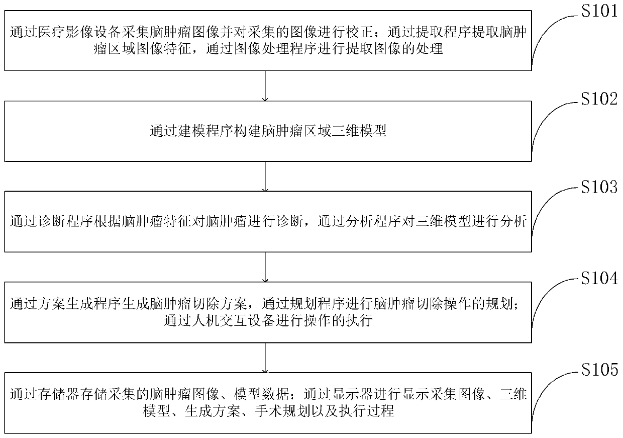
[0091] The three-dimensional reconstruction and display interaction method of brain tumor medical images provided by the embodiment of the present invention is as follows: figure 2 As shown, as a preferred embodiment, such as image 3 As shown, the method for correcting the collected image provided by the embodiment of the present invention includes the following steps:
[0092] S201, superimposing the collected brain tumor images to obtain a superimposed image;
[0093] S202. Linearly compress the superimposed image to obtain a normalized image;
[0094] S203. Determine the position and contour of the distorted image according to the pixel values of the normalized image at each position;
[0095] S204. Determine a correction factor of the distorted image, and determine a correction parameter;
[0096] S205. Determine a correction algorithm according to the correction parameter.
Embodiment 2
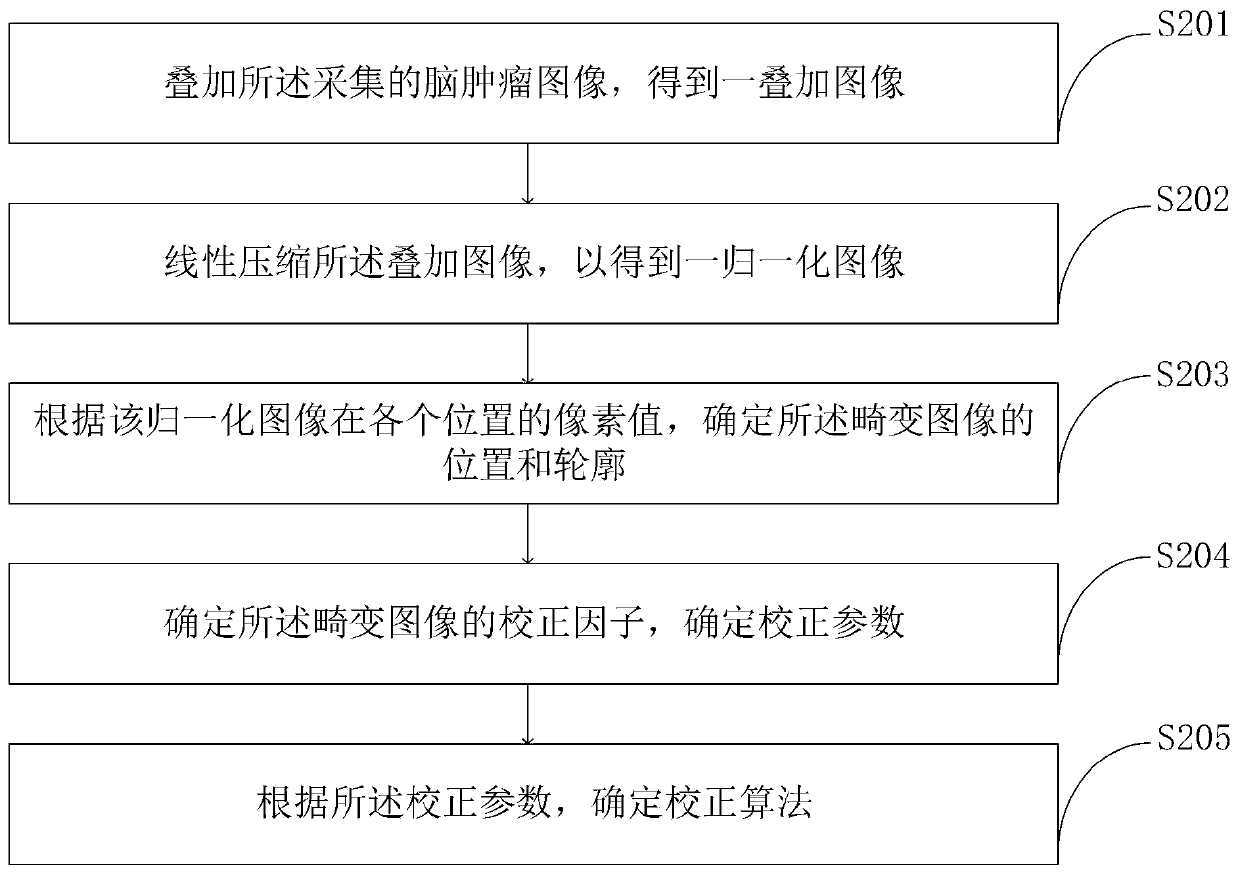
[0098] The three-dimensional reconstruction and display interaction method of brain tumor medical images provided by the embodiment of the present invention is as follows: figure 2 As shown, as a preferred embodiment, such as Figure 4 As shown, the method for constructing a three-dimensional model of a brain tumor region provided by an embodiment of the present invention includes the following steps:
[0099] S301. Based on the normalized plane profile of each image obtained in the image processing step, obtain the plane profile of each single three-dimensional object in three-dimensional space;
[0100] S302, based on the plane outlines of each single three-dimensional object in the three-dimensional space, splicing to obtain the multi-object plane outline in the three-dimensional space;
[0101] S303. Transform the spliced multi-object planar outlines in three-dimensional space into a multi-object 3D model.
Embodiment 3
[0103] The three-dimensional reconstruction and display interaction method of brain tumor medical images provided by the embodiment of the present invention is as follows: figure 2 As shown, as a preferred embodiment, such as Figure 5 As shown, the execution method of the operation provided by the embodiment of the present invention is:
[0104] S401, displaying the three-dimensional model of the brain tumor and the resection plan through the display screen;
[0105] S402, perform brain tumor location on the three-dimensional model, deduce the optimal operation plan and simulate the tumor resection process, the process is displayed on the display module and stored in the storage unit;
[0106] S403, use the data glove to build a virtual hand, and configure the virtual tool, combine the virtual hand and the virtual tool to simulate the resection operation on the three-dimensional model, simulate the feasibility of the surgical operation, and be proficient in the surgical pro...
PUM
 Abstract
Description
Claims
Application Information
 - Generate Ideas
- Intellectual Property
- Life Sciences
- Materials
- Tech Scout
- Unparalleled Data Quality
- Higher Quality Content
- 60% Fewer Hallucinations
Browse by: Latest US Patents, China's latest patents, Technical Efficacy Thesaurus, Application Domain, Technology Topic, Popular Technical Reports.
© 2025 PatSnap. All rights reserved.Legal|Privacy policy|Modern Slavery Act Transparency Statement|Sitemap|About US| Contact US: help@patsnap.com