Eureka AIR delivers breakthrough ideas for toughest innovation challenges, trusted by R&D personnel around the world.

Uterine contraction detection device based on maternal physiological electric signals and method thereof

A detection device and technology for electrical signals, which are used in pulse rate/heart rate measurement, diagnostic recording/measurement, medical science, etc., and can solve problems such as decreased algorithm accuracy, large amount of time-frequency domain analysis and calculation, and impact.

Active Publication Date: 2020-04-10
SOUTH SUZHOU MEDICAL TECH CO LTD
View PDF11 Cites 0 Cited by
  • Summary
  • Abstract
  • Description
  • Claims
  • Application Information

AI Technical Summary

Problems solved by technology

Time-frequency domain analysis can identify the burst band in the uterine myoelectric signal, and extract the uterine contraction wave signal corresponding to the burst wave, but it is sensitive to the selection of the calculation window length, and is easily affected by the large amplitude of the uterine myoelectric signal Impact of Impulse Noise
In addition, the time-frequency domain analysis has a large amount of calculation and is not suitable for real-time analysis systems
[0008] Nonlinear correlation coefficient H2, according to the nonlinear correlation coefficient of multiple leads to fit the uterine contraction curve, for the The noise suppression ability of a few leads with a large amplitude is strong, but the algorithm relies heavily on the acquisition system. When the number of leads in the acquisition system is small, the accuracy of the algorithm drops significantly
[0009]In short, the biggest challenge faced by the existing uterine contraction detection systems and algorithms is how to suppress the impact of the large noise in the EHG signal on the uterine contraction burst. At the same time, prevent the missed detection of some uterine contraction activities with small energy

Method used


Image

  • Uterine contraction detection device based on maternal physiological electric signals and method thereof
  • Uterine contraction detection device based on maternal physiological electric signals and method thereof
  • Uterine contraction detection device based on maternal physiological electric signals and method thereof

Examples


Embodiment 1

[0067] Step 1: Fusion of triaxial body position signals and RMS trend extraction

[0068] a) Fusion of three-axis acceleration signals:

[0069] The fusion of the three-axis body position signal is carried out according to the formula of formula (1):

[0070] acc(i)=|x t -x t-1 |+|y t -y t-1 |+|z t -z t-1 | (1)

[0071] x t 、y t ,z t Indicates the three-axis signal value collected by the acceleration sensor at time t, x t-1 、y t-1 ,z t-1 is the value at time t-1. The reason for using x t -x t-1 The form of this subtraction is because it is equivalent to a high-pass filter, which can remove the low-frequency noise introduced by the acceleration sensor due to breathing and other reasons.

[0072] b) The principle of the RMS algorithm and the extraction of body position trends:

[0073] The main principle of the RMS algorithm is to measure the energy change trend of the signal according to the standard deviation, as shown in formula (2):

[0074] The main princi...

Embodiment 2

[0122] Such as figure 2 As shown, a section of EHG signal (3 channels), MHR, body position signal and reference TOCO curve. As shown in the figure, it can be seen that when the uterine contraction occurs, the mother's heart rate fluctuates obviously, and the acceleration sensor also has obvious fluctuations. Variety.

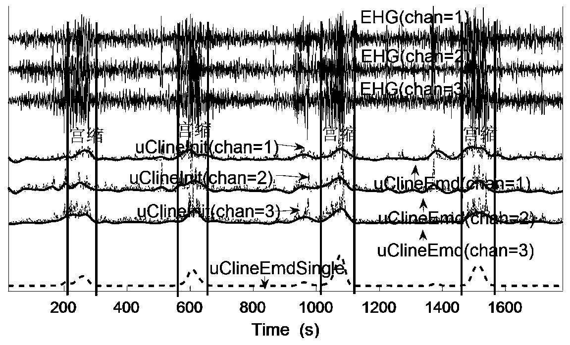
[0123] Such as image 3 As shown, a schematic diagram of an EHG signal (3 channels), RMS trend fitting (result is uClineInit), EMD optimization (result uClineEmd) and 3-channel uClineEmd fusion (result uClineEmdSingle).

[0124] Such as Figure 4 As shown, a section of EHG signal (3 channels) containing high and large pulse interference, EHG trend uClineEmdSingle extracted by this patent algorithm, and UC trend extracted only by RMS algorithm (N=60, M=3) are compared. It can be seen that in the noise section, the amplitude value corresponding to uClineEmdSingle obtained through EMD decomposition and other processing is significantly smaller than the amplitud...


PUM


Abstract

The invention discloses a uterine contraction detection device based on a maternal physiological electric signal and an acceleration sensor and a method thereof, and the device comprises an electrodewhich is in contact distribution with the abdominal body surface of a puerpera and obtains a body surface uterine myoelectric signal; a processor keeps electric signal transmission with the electrodes; the processor comprises a calculation and analysis system; the calculation and analysis system is used for extracting a uterine contraction trend curve from the body surface uterine electromyographysignals obtained by the electrodes and carrying out nonlinear decomposition and reconstruction on the uterine contraction trend curve through an EMD algorithm to obtain an accurate uterine contraction curve; and the scaling correction is carried on the uterine contraction accurate curve by integrating the maternal body position signal and the maternal heart rate data to obtain a final uterine contraction curve.

Description

technical field [0001] The invention relates to the field of medical appliances during pregnancy, in particular to a uterine contraction detection device and method based on maternal physiological electric signals and acceleration sensors. Background technique [0002] The current measurement methods of uterine contraction (UC) include TOCO (labor tonometry), IUP (intrauterine tonometry), and methods based on surface uterine electromyography (EHG). Among them, IUP is an invasive measurement method, which uses a catheter to measure intrauterine or amniotic fluid pressure, and may cause infection if it is not handled carefully. TOCO is non-invasive and is currently the most widely used clinical measurement method. During TOCO monitoring, a belt is wrapped around the abdomen of the parturient, and the uterine contraction can be felt by measuring the strain. The pressure measured in this way is seriously affected by the initial pressure of the measurement, and has a great relat...

Claims


Application Information

Patent Timeline
IPC IPC(8): A61B5/00A61B5/024A61B5/0488A61B5/11
CPCA61B5/4356A61B5/1116A61B5/024A61B5/7203A61B5/7225A61B5/389
Inventor 孙纪光
Owner SOUTH SUZHOU MEDICAL TECH CO LTD
Why Eureka
Eureka Blog
Learn More