
Blood cell segmentation and recognition model construction method and blood cell recognition method

A segmentation model and recognition model technology, applied in the field of medical images, can solve the problems of human factor interference, heavy workload, and low recognition accuracy, so as to reduce the interference of human objective factors, ensure accuracy and comprehensiveness, and improve objectivity. Effects of Sex and Consistency

Active Publication Date: 2020-01-03
BEIJING XIAOYING TECH CO LTD
View PDF4 Cites 8 Cited by
  • Summary
  • Abstract
  • Description
  • Claims
  • Application Information

AI Technical Summary

Problems solved by technology

[0002] The acquisition of whole slide electronic data is the basis for comprehensive and objective testing. The current medical inspection field, especially blood routine inspection, has heavy tasks and a heavy workload. A considerable number of hospitals have introduced advanced auxiliary inspection systems, but they cannot solve the problem of whole slide inspection. Inspection problems often lead to one-sided results and a high rate of manual re-examination; in addition, the serious shortage and uneven distribution of high-level laboratory physicians lead to different judgment results for abnormal cell morphology in peripheral blood. The current main identification and classification algorithms It belongs to the traditional sequence. In the actual operation process, the recognition accuracy is not high and it is easily interfered by subjective experience and human factors.
[0003] There are two main technical problems in the existing blood cell identification: (1) It is impossible to scan and analyze the whole field of view of the blood smear, resulting in one-sided and inaccurate results; As a result, it is extremely susceptible to interference from subjective experience and human factors, making the recognition accuracy low

Method used


Image

  • Blood cell segmentation and recognition model construction method and blood cell recognition method
  • Blood cell segmentation and recognition model construction method and blood cell recognition method
  • Blood cell segmentation and recognition model construction method and blood cell recognition method

Examples


Embodiment Construction

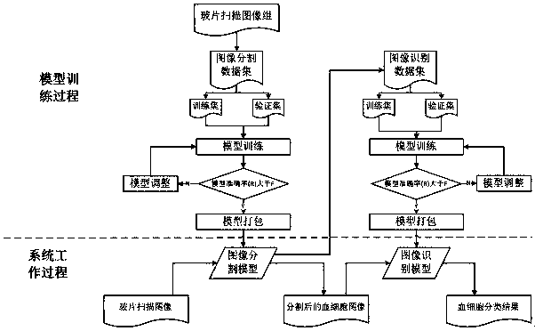
[0037] combine figure 1 On the one hand, a method for constructing a blood cell recognition model is provided to obtain a blood cell segmentation and recognition model for blood cell recognition. The specific steps are as follows:

[0038] (1) Image acquisition

[0039] Collect peripheral blood, make blood smears, digitize the collected blood samples and establish a blood image database, which stores full-slide full-field images of blood smears.

[0040] Due to the limited shooting range of the camera under a high-power microscope, especially under a 100X objective lens, it can only shoot a single-view image with a physical size of about 150*100μm (micrometer), such as Figure 5 As shown in (a) and (b), the blood cells at the edge of the single-view image cannot be accurately identified. In order to obtain images of the whole blood slide cells without omission (approximately 15mm*25mm in size), about 25,000 single-view images need to be stitched into a full-view image, such ...


PUM


Abstract

The invention relates to a blood cell segmentation and recognition model construction method and a blood cell recognition method, and provides a blood cell segmentation model and recognition model construction method on one hand and a cell recognition method based on a blood cell segmentation model and a recognition model on the other hand. The blood image database is generated based on the full-view image, and the blood cell segmentation model is trained, so that the accuracy and comprehensiveness of data are ensured, and the segmentation accuracy of the blood cell segmentation model is improved. Full-view blood cell analysis is realized by utilizing a computer, so that the interference of human objective factors is greatly reduced, and the objectivity and consistency of an inspection result are improved. The blood cell segmentation model and the recognition model are intelligent, the software algorithm has a self-learning attribute, the training efficiency of the recognition model isgradually improved along with the increase of high-quality labeled images, and the software recognition and classification accuracy can be continuously optimized.

Description

technical field [0001] The invention relates to a blood cell segmentation, a method for identifying a model structure and a blood cell identification method, belonging to the technical field of medical images. Background technique [0002] The acquisition of whole slide electronic data is the basis for comprehensive and objective testing. The current medical inspection field, especially blood routine inspection, has heavy tasks and a heavy workload. A considerable number of hospitals have introduced advanced auxiliary inspection systems, but they cannot solve the problem of whole slide inspection. Inspection problems often lead to one-sided results and a high rate of manual re-examination; in addition, the serious shortage and uneven distribution of high-level laboratory physicians lead to different judgment results for abnormal cell morphology in peripheral blood. The current main identification and classification algorithms It belongs to the traditional sequence. In the ac...

Claims


Application Information

Patent Timeline
IPC IPC(8): G06K9/00G06K9/62G06K9/34
CPCG06V20/695G06V10/267G06V2201/03G06F18/214
Inventor 方喆君李柏蕤连荷清吕东琦
Owner BEIJING XIAOYING TECH CO LTD
Learn More