
Neurosurgical postoperative drainage tube fixing device

A neurosurgery and fixation device technology, applied in catheters, suction devices, hypodermic injection devices, etc., can solve the problems of poor drainage, unsupported, troublesome nursing work, etc., to prevent entanglement, avoid mutual entanglement, and facilitate deployment. Effect

Pending Publication Date: 2019-12-20
AFFILIATED ZHONGSHAN HOSPITAL OF DALIAN UNIV
View PDF3 Cites 1 Cited by
  • Summary
  • Abstract
  • Description
  • Claims
  • Application Information

AI Technical Summary

Problems solved by technology

The main disadvantages of the above traditional methods are: first, the length of the neurosurgery drainage tube is generally about 180cm-200cm, and the drainage bag is only hung on the bottle holder and the drainage tube is left alone, which will easily cause the drainage tube to be entangled, discounted and the drainage is not enough. Second, many patients after neurosurgery are accompanied by agitation symptoms. When the drainage tube is not properly arranged or the patient's upper limbs are not properly restrained with a restraint belt, the patient will unconsciously pull the drainage tube on the head and easily pull out the drainage tube. Patient prognosis; third, it is not conducive to the implementation of clinical and nursing work. Some patients need to indwell two drainage tubes due to bilateral lateral ventricle hemorrhage or head tumors. The two drainage tubes are easily entangled with each other, which is not conducive to clinicians to identify or perform antibiotic flushing and drainage operations on them, and is also not conducive to the placement of pain relief pumps and depressurization pumps, which brings troubles to nursing work
The device can prevent the drainage tube from bending or kinking, but the telescopic rod of the device is prone to deformation during use, resulting in the inability to support the drainage tube on it and causing the entire device to fail

Method used


Image

  • Neurosurgical postoperative drainage tube fixing device
  • Neurosurgical postoperative drainage tube fixing device

Examples


Embodiment 1

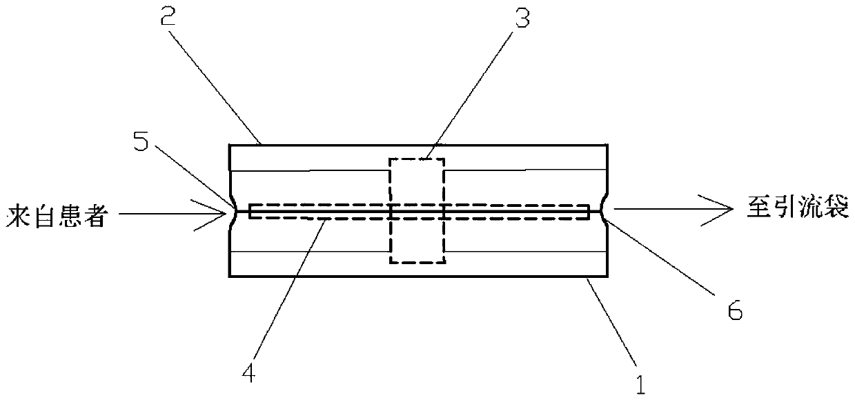
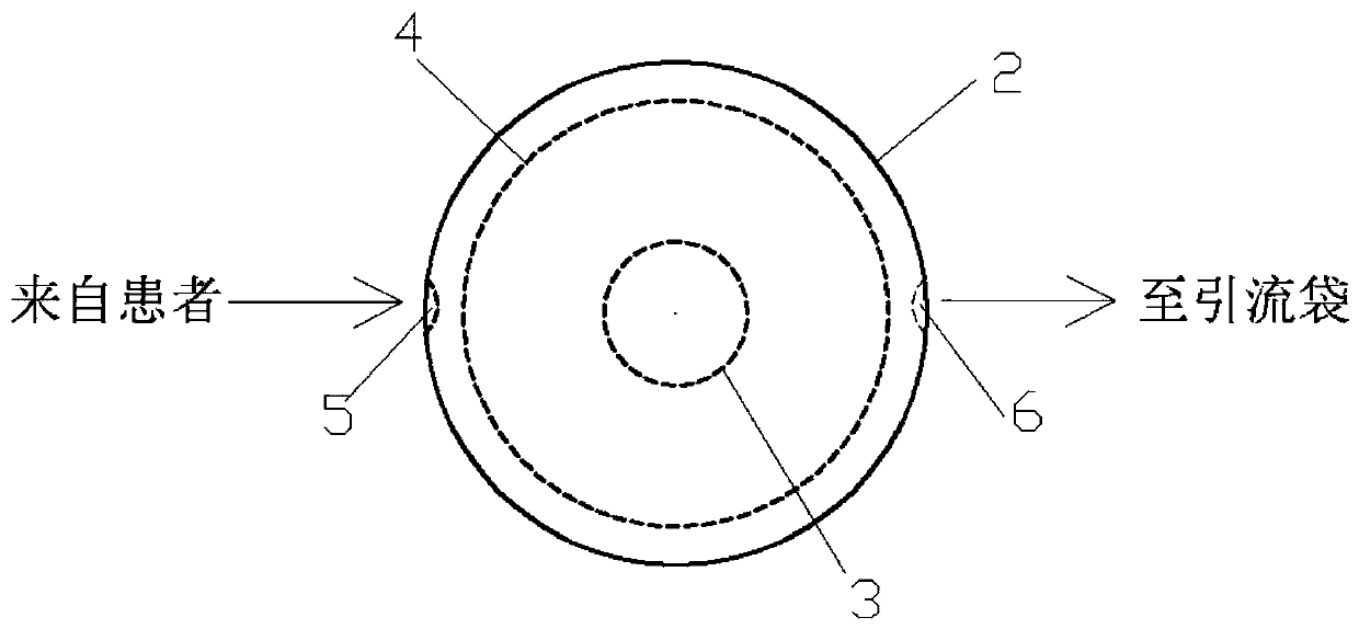
[0015] Example 1: See Figure 1-Figure 2 , the drainage tube fixing device after neurosurgery, consists of a cylindrical tray base 1, a cylindrical tray top cover 2, and a small cylinder 3 fixed inside the tray. Outer opening of the tray, the diameter of the tray base is 10cm, the diameter of the small cylinder is 7cm, the diameter of the tray is 10cm, and a disc 4 is embedded on the periphery of the small cylinder inside the tray, the diameter is 9.5cm, and the base 1 and the top cover 2 have The inlet circular hole 5 and the outlet circular hole 6, the inner diameters of the inlet circular hole 5 and the outlet circular hole 6 can meet the width of passing through two guide pipes at the same time, and the disc 4 divides the tray into upper and lower layers. There are small round holes on the outer edge of the tray 4 (not shown in the figure), and the inside of the cylindrical tray top cover 2 has threads, which can be matched and tightened with the threads of the tray base 1...


PUM


Abstract

The invention discloses a neurosurgical postoperative drainage tube fixing device. The device comprises a cylindrical tray base, a cylindrical tray top cover and a small cylinder which is fixed in a tray, a thread is arranged on the outer edge of the tray opening, a circle defined by the thread is called as a tray outer opening, a disc is embedded in the periphery of the small cylinder in the tray, inlet and outlet channels are formed in the base and the top cover, the disc divides the tray into an upper layer and a lower layer, small round holes are formed in the outer edge of the disc, and athread is arranged in the top cover, and the top cover can be screwed down on the tray base in thread connection. The device has the beneficial effects of: preventing the unsmooth drainage caused bywinding and folding of a drainage tube of a neurosurgical postoperative patient; effectively preventing the drainage tube from being unconsciously dragged and pulled out by the patient to influence prognosis in that the drainage tube is too long; and being more beneficial to implementation of clinical and nursing work, preventing drainage tubes from being intertwined when two drainage tubes need to be placed into a patient according to the illness state, and helping clinicians to conveniently recognize the drainage tubes or conduct antibiotic flushing, drainage and other operations on the drainage tubes.

Description

technical field [0001] The invention belongs to the field of surgical supplies, in particular to a drainage tube fixing device after neurosurgery. Background technique [0002] Common external drainage tubes in neurosurgery include subcutaneous drainage tubes, epidural drainage tubes, subdural drainage tubes, subarachnoid drainage tubes (lumbar cistern drainage tubes), lateral ventricular external drainage tubes, and wound (tumor) cavity drainage tubes. . [0003] The purpose of subcutaneous and epidural drainage tubes is mainly to prevent subcutaneous and epidural hematoma after craniotomy, and subdural drainage tube is mainly used for chronic subdural effusion or subdural hematoma because a complete capsule has been formed After surgery, the drainage tube was connected to the capsule to continue drainage to empty the residual bloody fluid or blood clot in the capsule, so as to facilitate the swelling of the brain tissue and eliminate the dead space. Lumbar drainage tubes...

Claims


Application Information

Patent Timeline
Patent Type & Authority Applications(China)
IPC IPC(8): A61M25/02A61M1/00
CPCA61M25/02A61M2025/028A61M1/84
Inventor 刘权邓东风张林林张绪新李彦钊李凯
Owner AFFILIATED ZHONGSHAN HOSPITAL OF DALIAN UNIV
Learn More