Detection of nerves in a series of echographic images

A series of technologies in images, applied in image enhancement, image analysis, image data processing, etc., can solve problems such as difficulties in automatic detection process

Inactive Publication Date: 2019-12-13
UNIV OF ORLEANS +1
View PDF2 Cites 0 Cited by
  • Summary
  • Abstract
  • Description
  • Claims
  • Application Information

AI Technical Summary

Problems solved by technology

The variability of neural structures means that their appearance on images is also highly variable and makes the automatic detection process even more difficult

Method used


Image

  • Detection of nerves in a series of echographic images
  • Detection of nerves in a series of echographic images
  • Detection of nerves in a series of echographic images

Examples


Embodiment Construction

[0026] In practice, the anesthesiologist scans areas of the patient's body with an ultrasound probe to identify nerves and determine the areas where the anesthetic product is to be injected. The ultrasound device operates continuously under the control of the anesthesiologist or another practitioner (surgeon, etc.) and generates a stream of images (ie, video) that are displayed on a monitor in real time. Thereby, scans performed by the anesthetist can be undertaken based on what the anesthetist sees on the monitor.

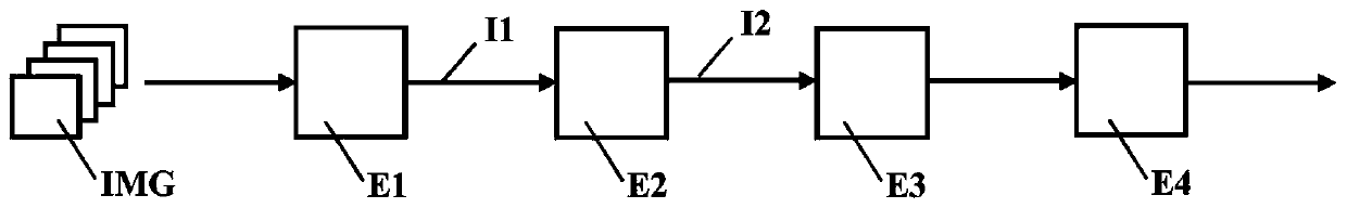
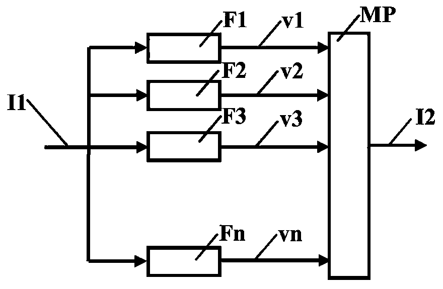
[0027] The method according to the invention is based on a processing chain which acquires as input a stream of echographic images and supplies as output the detection of nerves on this stream.

[0028] Typically, the detection may include highlighting the nerve as overexposed on the stream of echographic images displayed on the monitor. For example, such protrusions may include enclosing the detected nerve or even precisely defining a contour. It can be done in...


PUM


Abstract

The invention concerns a method for detecting a nerve in a series of echographic images comprising, for each image of said series: a step (E2) of generating a map of probabilities over regions of theimage, consisting of applying a plurality of texture descriptors in order to generate, for each pixel, a vector determining the response of the pixel for each of the descriptors, and then deducing a probability for each pixel of belonging to a nerve as a function of a probabilistic model; a classification step (E3) applied on the zones determined by this probability map, consisting of searching models corresponding to a nerve type, in a window sliding over these zones, assigning a degree of confidence to each position of the window and retaining an optimum position for each model; then analysing the consistency of these optimum positions by measuring their stability over a set of images of the series in order to select the window having the maximum consistency, which corresponds to a detected nerve.

Description

technical field [0001] The invention relates to the field of analysis of digital images derived from ultrasound scans. More specifically, the invention relates to the automatic detection of nerves in a series of images to assist anesthesiologists in their work. Background technique [0002] Local regional anesthesia involves injecting an anesthetic product near a patient's nerves. Therefore, it is important that anesthesiologists have tools that facilitate their work by allowing them to precisely locate nerves. To this end, anesthesiologists have equipment that allows them to observe in real time an echographic image of the anatomical region of the patient under study. An anesthesiologist can scan the area to locate nerves and determine suitable locations for placing anesthesia products. [0003] However, detecting nerves in echographic images is a difficult task even for experienced anesthesiologists. [0004] Therefore, it is of interest to have anesthesiologists with ...

Claims


Application Information

Patent Timeline
Patent Type & Authority Applications(China)
IPC IPC(8): G06K9/00G06T7/00G06T7/41G06T7/73G06T7/77
CPCG06T7/0012G06T2207/10016G06T2207/10132G06T2207/20064G06T2207/20081G06T7/75G06T7/77G06T7/41G06T2207/20084G06V40/10G06V40/14G06V10/467G06T2207/30024
Inventor 阿德尔·哈菲亚奈乌萨马·哈吉赫西皮埃尔·维雷斯伊夫·帕尔芒捷
Owner UNIV OF ORLEANS
Why Eureka
Try Eureka