
A method for preparing neutrophil microvesicles

A neutrophil and microvesicle technology, applied in biochemical equipment and methods, animal cells, vertebrate cells, etc., can solve the problems of undiscovered research reports and few studies on neutrophil microvesicles, etc. The effect of preventing excessive damage and uniform size

Active Publication Date: 2020-07-17
海南省妇幼保健院
View PDF2 Cites 0 Cited by
  • Summary
  • Abstract
  • Description
  • Claims
  • Application Information

AI Technical Summary

Problems solved by technology

Studies have shown that neutrophils have a certain role in the treatment of arthritis. In addition, there are relatively few studies on neutrophil microvesicles, especially the relationship between the diameter of microvesicles and their biological functions. relationship, no relevant research reports have been found

Method used


Image

  • A method for preparing neutrophil microvesicles
  • A method for preparing neutrophil microvesicles

Examples


Embodiment 1

[0023] A method for preparing neutrophil microvesicles, comprising the following steps:

[0024] S1: Add the stimulator to the neutrophil culture medium (the cell concentration is 10 5 cell / mL), let stand for 45min, UVB radiation (radiation energy is 10mJ / cm 2 )30min;

[0025] S2: Add 33 mg / L folic acid (that is, add 33 mg folic acid per liter of solution), take the cell culture solution after 3 hours, centrifuge at 2000×g, 0°C for 3 minutes, collect the supernatant, and add chlorogenic acid to the supernatant (per liter of folic acid). Add 12 mg) to the supernatant, centrifuge at 6000×g, 0°C for 25 min after 15 min, discard the supernatant, and obtain neutrophil microvesicles;

[0026] Among them, before centrifugation, always keep the solution temperature at 15-18°C;

[0027] The stimulating agent is a mixture of calcium chloride, folic acid, glucose and proanthocyanidins (mass ratio is 1:0.1:20:0.1);

[0028] The dosage of the stimulating agent is 0.5g / L neutrophil cult...

Embodiment 2

[0031] A method for preparing neutrophil microvesicles, comprising the following steps:

[0032] S1: Add the stimulator to the neutrophil culture medium (the cell concentration is 10 7 cell / mL), let stand for 30min, UVB radiation (radiation energy is 15mJ / cm 2 )15min;

[0033] S2: Add 38mg / L folic acid, take the cell culture fluid after 2h, centrifuge at 3000×g, 4°C for 5min, collect the supernatant, add chlorogenic acid (20mg per liter of supernatant) to the supernatant, 10min Afterwards, centrifuge at 8000×g, 4°C for 40 minutes, discard the supernatant, and obtain neutrophil microvesicles;

[0034] Among them, before centrifugation, always keep the solution temperature at 15-18°C;

[0035] The stimulating agent is a mixture of calcium chloride, folic acid, glucose and proanthocyanidins (the mass ratio of calcium chloride, folic acid, glucose and proanthocyanidins is 1:0.2:28:0.2);

[0036] The consumption of stimulating agent is 1.0g / L neutrophil culture medium;

[0037...

Embodiment 3

[0039] The difference between embodiment 3 and embodiment 1 is:

[0040] Step S2 is:

[0041] Add 33 mg / L folic acid, take the cell culture medium after 3 hours, centrifuge at 6000×g, 0°C for 25 minutes, discard the supernatant, and obtain neutrophil microvesicles.


PUM

PropertyMeasurementUnit
diameteraaaaaaaaaa
diameteraaaaaaaaaa

Abstract

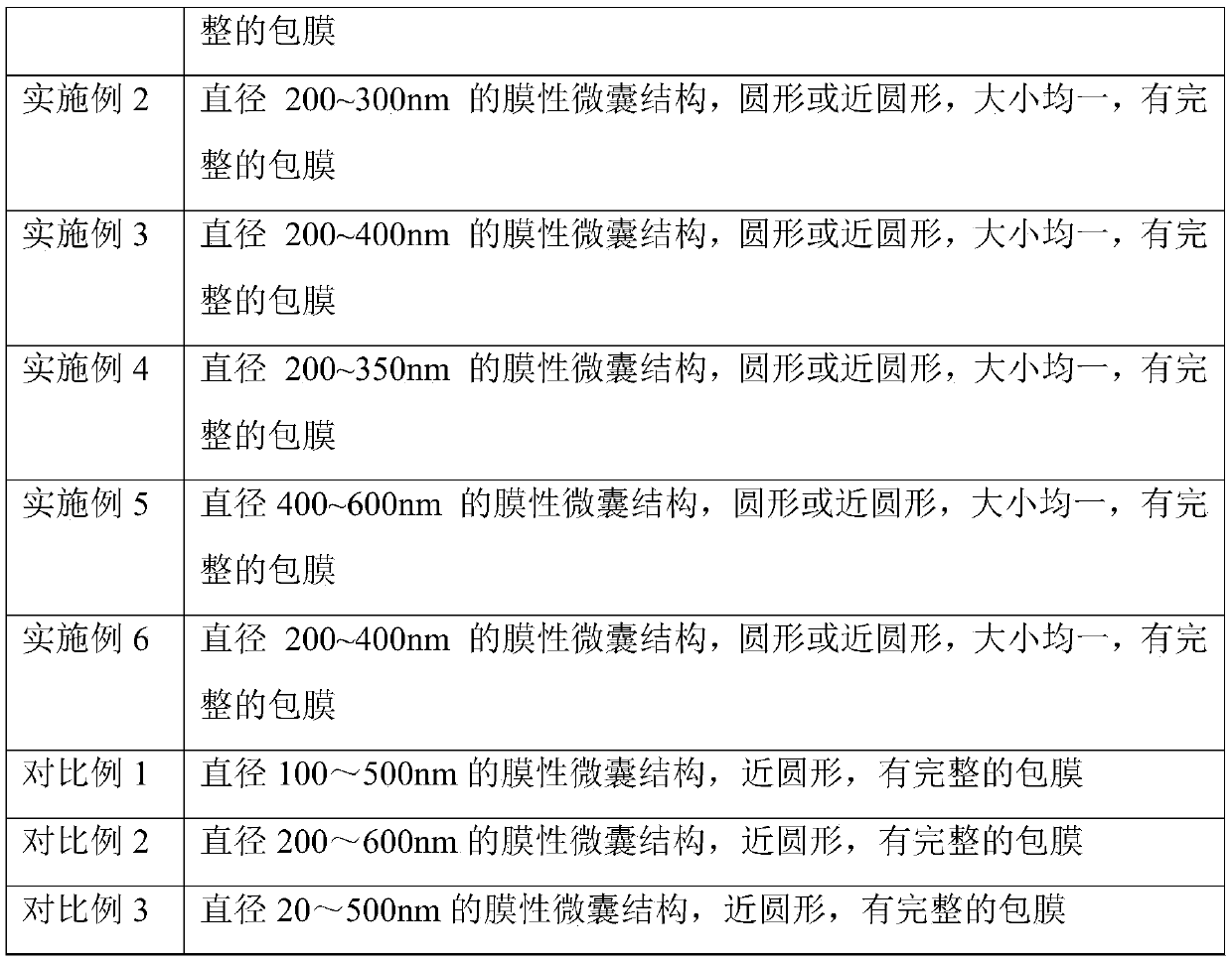
The invention discloses a method for preparing neutrophil microvesicles, which comprises the following steps: adding a stimulant to a neutrophil culture medium, performing standing for 30 to 45 min, and irradiating the material under ultraviolet rays for 15 to 30 min; and adding folic acid, and collecting the neutrophil microvesicles by centrifugation after 2 to 3 hours, wherein the solution temperature is kept at 15 to 18 DEG C before centrifugation, and the stimulant is a mixture of calcium chloride, folic acid, glucose and procyanidin. The method of the invention can rapidly stimulate the neutrophils to produce microvesicles with uniform size, while maintaining the normal growth state of the cells and preventing excessive damage to the cells. By adopting the method of the invention, thedifference in diameter of the obtained neutrophil microvesicles does not exceed 200 nm, the size is uniform, and a complete envelope is provided.

Description

technical field [0001] The invention relates to a method for preparing neutrophil microvesicles, belonging to the field of biotechnology. Background technique [0002] Microvesicles (MVs) are the first cell membrane-derived vesicle structures discovered in the blood system, with a diameter usually between 100-1000 nm (some scholars believe that the diameter is between 20-1000 nm). Studies have found that a variety of cells can produce microvesicles, such as epithelial cells, fibroblasts, immune cells, and tumor cells. Neutrophils are the body's first barrier against microorganisms and play an important role in the natural immune system. The microvesicles released by them contain protein and lipid components similar to those of mother cells, and may also contain mother cell cytoplasm Therefore, neutrophil microvesicles may participate in the transmission of information between cells, participate in inflammatory responses, immune responses, etc. Studies have shown that neutr...

Claims


Application Information

Patent Timeline
Patent Type & Authority Patents(China)
IPC IPC(8): C12N5/0787C12N13/00
CPCC12N5/0642C12N13/00C12N2500/14C12N2500/34C12N2500/38C12N2501/999
Inventor 战跃福吴烨华王毅翔鲁宏郭子义陈建强关莹
Owner 海南省妇幼保健院
Learn More