
Video-assisted endoscopic set for anal fistula treatment

A technology of endoscopy and video, applied in the field of video-assisted endoscopy kits, to achieve good surgical results, save time, and easy operation

Pending Publication Date: 2019-09-20
YUEYANG INTEGRATED TRADITIONAL CHINESE & WESTERN MEDICINE HOSPITAL SHANGHAI UNIV OF CHINESE TRADITIONAL MEDICINE
View PDF6 Cites 8 Cited by
  • Summary
  • Abstract
  • Description
  • Claims
  • Application Information

AI Technical Summary

Problems solved by technology

[0004] However, there is currently no video-assisted endoscopy kit suitable for the treatment of anal fistula, especially the dual functions of visual exploration of fistula and laser treatment, no need to replace instruments during the operation, easy operation, time-saving, and good therapeutic effect of video-assisted endoscopy mirror kit device

Method used


Image

  • Video-assisted endoscopic set for anal fistula treatment
  • Video-assisted endoscopic set for anal fistula treatment
  • Video-assisted endoscopic set for anal fistula treatment

Examples


Embodiment 1

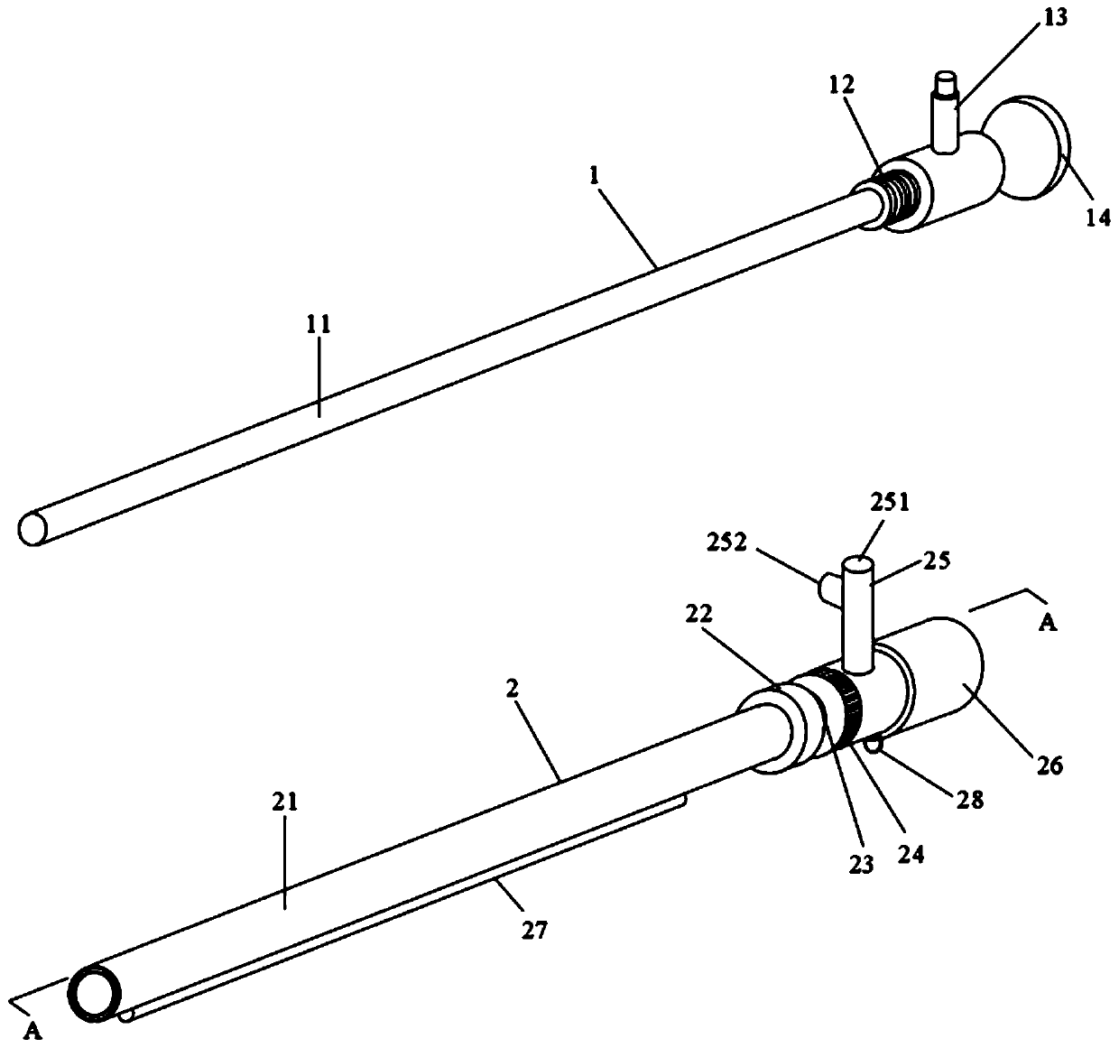
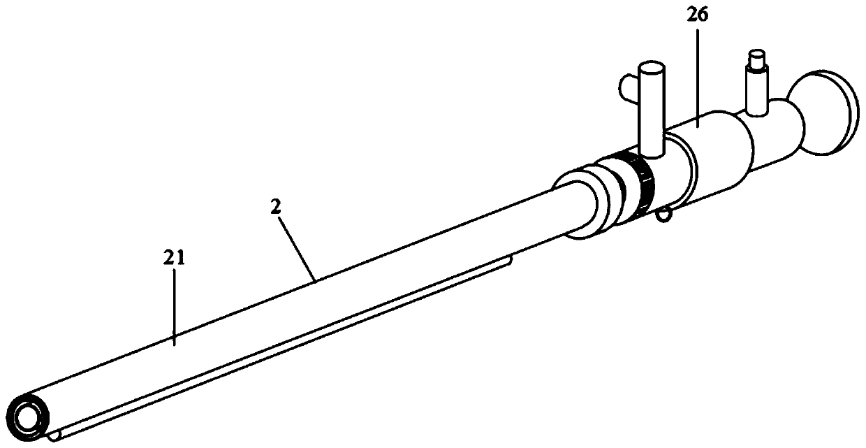
[0046] See figure 1 , figure 1 It is a structural schematic diagram of the disassembled state of the video-assisted endoscope set for anal fistula treatment according to the present invention. The video-assisted endoscope kit for anal fistula treatment is provided with an endoscope 1 and an outer sheath 2 . The endoscope 1 is sequentially provided with a mirror body 11 , a first assembly joint 12 , a light source joint 13 and an image equipment joint 14 from the distal end to the proximal end. The mirror body 11 is long cylindrical, and the first assembly joint 12 is provided with external threads. The outer sheath 2 is sequentially provided with an endoscope sleeve 21 , a stop ring 22 , a slideway 23 , a push-pull ring 24 , a tee 25 and a second assembly joint 26 from the distal end to the proximal end. The endoscope sleeve 21 is a long tubular body with a hollow interior, and the outer wall of the endoscope sleeve 21 is provided with an optical fiber operation channel 27....

Embodiment 2

[0058] See Figure 5 , Figure 5 It is a structural schematic diagram of the video-assisted endoscope system for anal fistula treatment according to the present invention. The video-assisted endoscope system includes the video-assisted endoscope kit 100 for anal fistula treatment as described in Embodiment 1, and also includes a laser therapy instrument 200 , an imaging device 300 and a light source device 400 . The laser therapy instrument 200 is provided with an optical fiber, the image device 300 is connected to the image device connector 14 of the video-assisted endoscope kit 100 for anal fistula treatment, and the light source device 400 is connected to the video-assisted endoscope set 100 for anal fistula treatment. The light source connector 13 of the mirror assembly 100 is connected.


PUM

PropertyMeasurementUnit
Lengthaaaaaaaaaa
Outer diameteraaaaaaaaaa
Lengthaaaaaaaaaa

Abstract

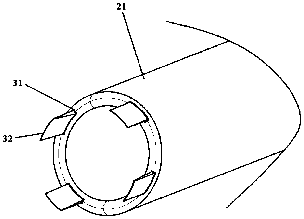
The invention relates to a video-assisted endoscopic set for anal fistula treatment. The video-assisted endoscopic set comprises an endoscope and an outer sheath, the endoscope comprises an endoscope body, a first assembly connector, a light source connector and an image device connector, and the outer sheath comprises an endoscope sleeve, a slide way, a push-pull ring, a tee joint, a second assembly connector and an optical fiber operating passage. The side wall of the endoscope sleeve is provided with a blade groove, and the far end of the blade groove extends to a far end face of the endoscope sleeve while the near end extends to communicate with the slide way. A blade and a push-pull rod are arranged in the blade groove, the near end of the push-pull rod extends out of the blade groove to connect with the push-pull ring, and the push-pull ring is capable of moving along the slide way to enable the blade to extend out of or hide in the endoscope sleeve. The endoscope sleeve externally sleeves the endoscope body, and the first assembly connector and the second assembly connector are fastened. The video-assisted endoscopic set is applied to anal fistula surgery, fistula probing and treatment can be carried out under direct vision and can be completed by one step, instrument changing is avoided during the surgery, narrow or adhered fistulae can be treated conveniently, and high surgical efficiency and effectiveness are achieved.

Description

technical field [0001] The invention relates to the technical field of medical instruments, in particular to a video-assisted endoscope set for anal fistula treatment. Background technique [0002] Anal fistula is mostly caused by infection and suppuration of the perianal space. After abscess incision and drainage or spontaneous ulceration, the abscess cavity shrinks and the inner wall becomes fibrotic. It consists of a primary internal opening, a pathological fistula tract, and a secondary external opening. Accurate detection of the fistula tract and treatment of the primary internal opening are the keys to curing anal fistula. In clinical treatment, due to the complexity of anal fistula disease itself, for the presence of multiple fistulas at the same time, fistulas with deep or arc-shaped fistulas, and high-level complex anal fistulas with fistulas deep in the anal sphincter, clinical treatment trickier. Modern medicine is based on the principle of surgical treatment of...

Claims


Application Information

Patent Timeline
IPC IPC(8): A61B1/31A61B1/018A61B1/015A61B1/04A61B1/06A61B17/3209A61B18/22
CPCA61B1/31A61B1/018A61B1/015A61B1/04A61B1/06A61B1/00071A61B1/00131A61B17/3209A61B17/320016A61B18/22A61B2018/2205A61B2018/005A61B2018/00982
Inventor 吴炯王振宜金磊杨豪杰秦凯健崔灿吴人杰许愿许曼
Owner YUEYANG INTEGRATED TRADITIONAL CHINESE & WESTERN MEDICINE HOSPITAL SHANGHAI UNIV OF CHINESE TRADITIONAL MEDICINE
Learn More