
Lung CT image adhesion vascular nodule detection method

A technology of CT image and detection method, applied in the field of CT image detection and processing, to achieve the effect of concise design idea, reduction of false positives, and small amount of calculation

Inactive Publication Date: 2019-09-10
JILIN UNIV FIRST HOSPITAL
View PDF3 Cites 3 Cited by
  • Summary
  • Abstract
  • Description
  • Claims
  • Application Information

AI Technical Summary

Problems solved by technology

[0003] The technical problem to be solved by the present invention is to provide a method for detecting adherent vascular nodules in lung CT images to solve the defects in the prior art

Method used


Image

  • Lung CT image adhesion vascular nodule detection method

Examples


Embodiment Construction

[0015] The principles and features of the present invention are described below in conjunction with the accompanying drawings, and the examples given are only used to explain the present invention, and are not intended to limit the scope of the present invention.

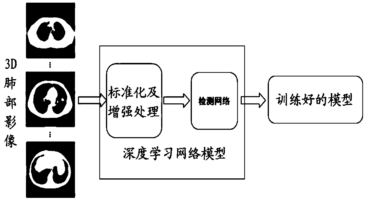
[0016] Such as figure 1 As shown, a deep learning-based CT pulmonary nodule detection method includes the following steps:

[0017] Step 1: Obtain the user's 3D lung CT sequence image;

[0018] Step 2: Standardize the 3D image to obtain multiple 3D cube sample blocks of the same size;

[0019] Step 3: Enhance the standardized 3D image data, and then input it to the preset deep learning network model for training, so as to obtain a trained pulmonary nodule detection model;

[0020] Step 4: Input the tested 3D lung CT sequence images into the trained pulmonary nodule detection model to obtain preliminary pulmonary nodule detection results;

[0021] Step 5: For the preliminary pulmonary nodule detection results, the...


PUM


Abstract

The invention relates to a lung CT image adhesion vascular nodule detection method. The method comprises the following steps: 1, obtaining a 3D lung CT sequence image of a user; 2, standardizing the 3D image to obtain a plurality of 3D cube sample blocks with the same size; 3, performing enhancement processing on the standardized 3D image data, and then inputting the standardized 3D image data into a preset deep learning network model for training, thereby obtaining a trained pulmonary nodule detection model; 4, inputting the tested 3D lung CT sequence image into the trained pulmonary nodule detection model so as to obtain a preliminary pulmonary nodule detection result; and 5, for a preliminary pulmonary nodule detection result, drawing an ROC curve according to a result of the test sample set, and determining an optimal threshold value according to characteristics of the ROC curve and an AUC value for classifying benign and malignant pulmonary nodules by using the trained new neuralnetwork model. Under the condition that the overall precision is guaranteed, false positive can be further reduced, the film reading pressure of doctors is greatly reduced, the doctors can be more focused on other more creative tasks, and huge economic and social benefits are achieved.

Description

technical field [0001] The invention relates to the technical field of CT image detection and processing, in particular to a method for detecting adherent vascular nodules in lung CT images. Background technique [0002] Lung cancer is the main cause of cancer-related death worldwide. Using CT scans to check high-risk groups is an effective means of detecting early lung cancer. Early detection of lung nodules is the key to improving the survival rate of lung cancer patients. The discovery of pulmonary nodules is the first step in the prevention and treatment of early lung cancer. With the advent of the era of big data, hospitals will generate a large amount of CT image data every day, which puts enormous pressure on radiologists to read images. According to statistics, when doctors read more than 20 sets of films per day, the error rate will reach 7%-15%. Therefore, it is of great significance to develop an automatic detection method for pulmonary nodules to improve the wo...

Claims


Application Information

Patent Timeline
Patent Type & Authority Applications(China)
IPC IPC(8): G06T7/00
CPCG06T7/0012G06T2207/10081G06T2207/30064G06T2207/20081G06T2207/20084
Inventor 华树成
Owner JILIN UNIV FIRST HOSPITAL
Learn More