Method for constructing model of huge mass in uterus and fetal neck

A fetus and uterus technology, applied in the field of medical application research, can solve the problem of difficulty in simulating patients with fetal neck mass, and achieve the effects of dustproof and waterproof, prolonging service life and enhancing accuracy

Inactive Publication Date: 2019-08-30
THE WEST CHINA SECOND UNIV HOSPITAL OF SICHUAN
View PDF6 Cites 2 Cited by
  • Summary
  • Abstract
  • Description
  • Claims
  • Application Information

AI Technical Summary

Problems solved by technology

It is difficult for general medical courses to simulate real patients with fetal neck masses. The complex characteristics of 3D printing materials and the ability to easily create models in 3D make it possible to develop realistic models based on real uterus and patients with huge fetal neck masses.
[0005] In the prior art, there is not a complete set of construction methods for constructing a large mass model of the uterus and fetal neck. Therefore, it is urgent to design a method for constructing a large mass model of the uterus and fetal neck, so that it can be conveniently Obstetricians and anesthesiologists can formulate safe and reliable clinical diagnosis and treatment plans for such patients based on the constructed uterine and fetal neck huge mass models, which will help the training and teaching demonstration of clinical skills for such diseases, and improve the Clinical Practice Skills for Senior Clinicians

Method used


Image

  • Method for constructing model of huge mass in uterus and fetal neck

Examples


Embodiment

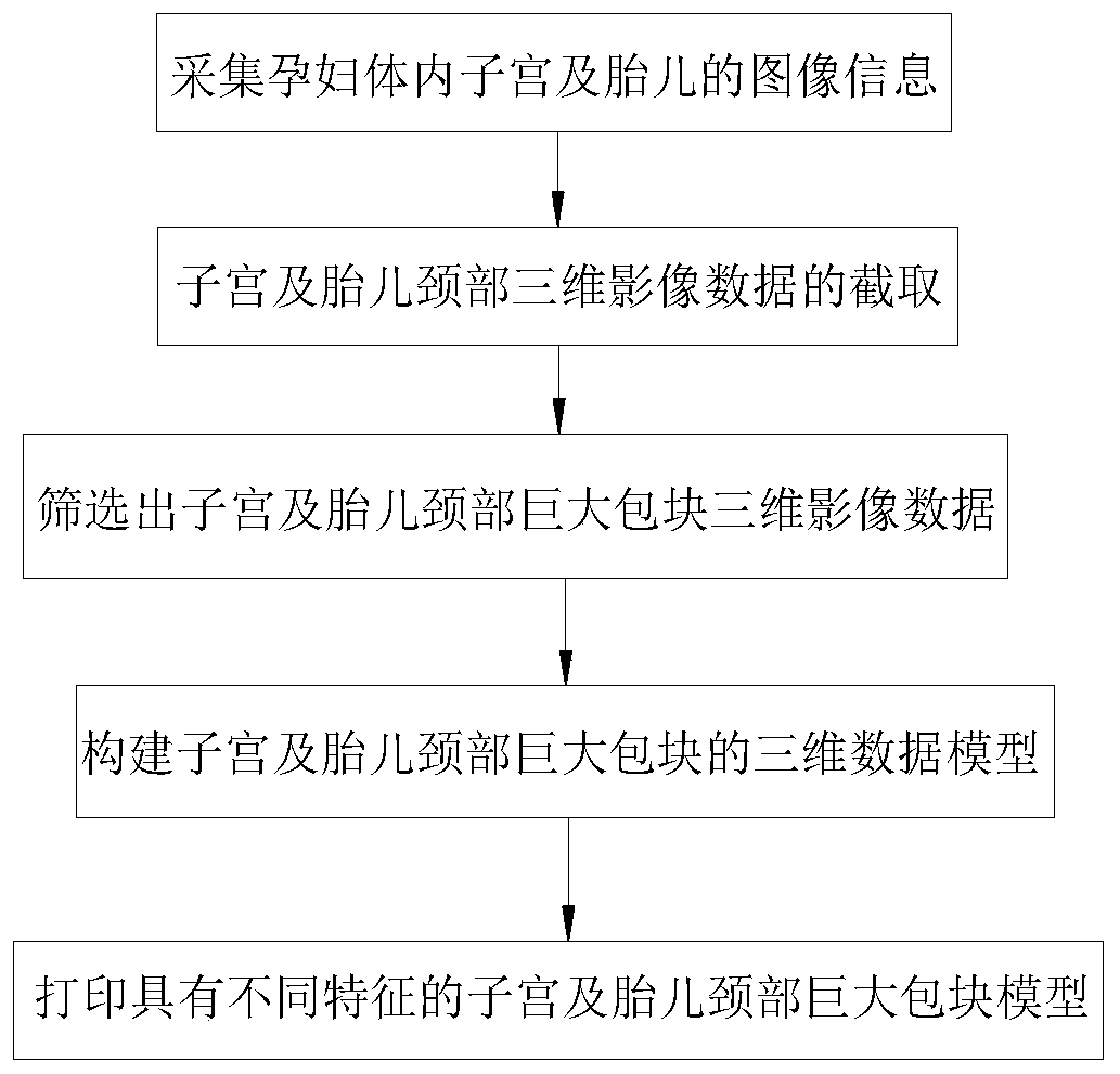
[0027] Example: A method for constructing a huge mass model of the uterus and fetal neck, such as figure 1 As shown, including the following steps:

[0028] 1) Image information acquisition: Scan and image the fetus in the pregnant woman through the scanning imaging device, and transfer the image information data collected by the scanning imaging device to the data processing terminal communicating with the scanning imaging device for processing and storage, to obtain the uterus and Three-dimensional image data of the fetus.

[0029] 2) According to the three-dimensional image data of the uterus and fetus obtained in step 1), the three-dimensional image data of the uterus and the fetal neck are intercepted through the three-dimensional image data cutting software implanted in the data processing terminal.

[0030] 3) According to the three-dimensional image data of the uterus and fetal neck intercepted in step 2), the three-dimensional image data of the uterus and fetal neck is inte...


PUM


Abstract

The invention discloses a method for constructing a model of a huge mass in a uterus and a fetal neck, and relates to the technical field of medical application research. According to the key points of the technical scheme, the method comprises the following steps that (1) image information collection is performed; (2) three-dimensional image data of the uterus and the fetal neck are intercepted;(3) the three-dimensional image data of a huge mass in the uterus and fetal neck are judged and screened out; (4) three-dimensional models of the huge mass in the uterus and fetal neck with differentcharacteristics are constructed through a three-dimensional model building system; and (5) a plurality of uterus and fetal neck huge mass models with different characteristics are printed through a 3Dprinting device. The method has the effects that a solid model of the huge mass in the uterus and the fetal neck can be constructed, doctors can develop a safe and reliable clinical diagnosis and treatment plan for patients conveniently according to the model of the huge mass in the fetal neck, the training of clinical skills of the disease and teaching display are facilitated by the model, and the clinical practice skills of students and junior clinicians are improved conveniently.

Description

Technical field [0001] The invention relates to the technical field of medical application research, more specifically, it relates to a method for constructing a huge mass model of the uterus and fetal neck. Background technique [0002] Although the incidence of serious fetal neck masses such as lymphatic cysts, pharyngeal neck teratomas, and cervical hemangioma is low, if not handled properly, it can lead to serious consequences. Severe huge neck mass can grow to the mediastinum, causing severe airway compression of the fetus. Failure of tracheal intubation may occur when the fetus is born, leading to hypoxia, brain damage, and even death. The early diagnosis of these diseases during the fetal period and the prevention and treatment of failure of tracheal intubation at birth are very important, and directly determine the recovery of the fetus. [0003] At present, the research data on the huge mass of the fetal neck is mainly carried out through the image data obtained by medic...

Claims


Application Information

Patent Timeline
IPC IPC(8): G09B23/28
CPCG09B23/281
Inventor 江晓琴童煜马玉姗冯世苗宋丽平吴兰康炎茗林雪梅
Owner THE WEST CHINA SECOND UNIV HOSPITAL OF SICHUAN
Why Eureka
Try Eureka