Preparation of protein micro crystal frozen sample and method of structural analysis

An analytical method and crystal structure technology, applied in the field of structural biology, can solve problems such as the inability to resolve atomic resolution structures of tiny crystals

Pending Publication Date: 2019-08-06
SHANGHAI INST OF ORGANIC CHEM CHINESE ACAD OF SCI
View PDF4 Cites 0 Cited by
  • Summary
  • Abstract
  • Description
  • Claims
  • Application Information

AI Technical Summary

Problems solved by technology

However, many important proteins, such as pathogenic amyloid and membrane proteins in neurodegenerative diseases, can only form ultrafine crystals of tens of nanometers to several micrometers due to the influence of many factors such as their own flexibility and heterogeneity. X-ray diffraction technology cannot analyze the atomic resolution structure of such tiny crystals, so a cryo-electron microscopy electron diffraction method that is feasible for this type of crystals is invented to analyze the atomic resolution structure of such tiny crystals
However, microcrystal electron diffraction technology is a novel technology, and there is no mature method for sample preparation and structure analysis of tiny crystals of proteins.

Method used


Image

  • Preparation of protein micro crystal frozen sample and method of structural analysis
  • Preparation of protein micro crystal frozen sample and method of structural analysis

Examples


Embodiment 1

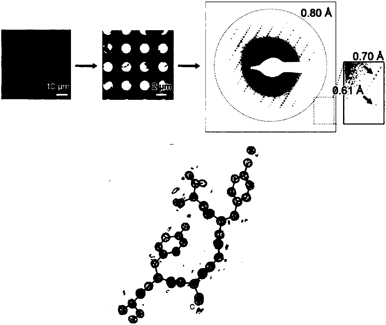
[0082] Structural Analysis of Peptide SYSGYS

[0083] 1) Sample processing

[0084] First use a needle-shaped tool to smash the middle nucleus, so that the needle-shaped crystals can be evenly dispersed in the droplet, then suck the crystals together with the pool liquid into the centrifuge tube, and dilute appropriately to obtain a suspension of crystals, use Prepared for frozen samples.

[0085] 2) Frozen sample preparation

[0086] The sample volume was determined according to the concentration of the crystals, the volume was 4 μl, and the sample preparation was performed manually and in conjunction with Vitrobot. First, load an appropriate amount of sample onto the hydrophilized frozen copper grid. Generally, Quantifoil’s model is 2 / 2, 400 mesh copper grid. Copper grid. If the crystal concentration is low, the sample can be loaded multiple times and blotted dry on the reverse side of the copper grid. After absorbing the liquid, wash it with 4 μl 5% PEG200 solution, an...

Embodiment 2

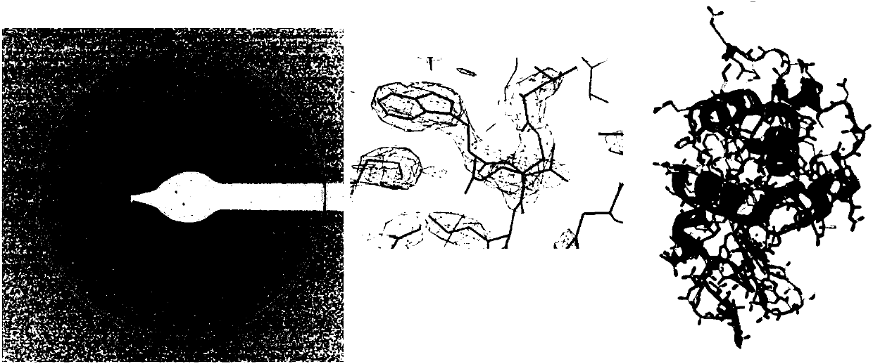
[0096] Elucidation of the regular crystal structure of lysozyme.

[0097] 1) Sample processing

[0098] Transfer an appropriate amount of sample and crystallization solution to an appropriate size ep tube. Water bath ultrasound: Ultrasonic in the water bath ultrasonicator adjusted to the lowest power, the ultrasonic power is 20%, the water bath time is 0.5s, try not to damage the integrity of the crystal package. After ultrasonication, spin the sample so that large pieces are deposited at the bottom of the test tube, and gently take the upper sample for sample preparation.

[0099] The steps of frozen sample preparation, crystal screening, data collection and structure analysis are the same as in Example 1.

[0100] Parsing results such as figure 2 As shown, the resolution of the conventional crystal structure of the precipitated lysozyme is


PUM

PropertyMeasurementUnit
thicknessaaaaaaaaaa
thicknessaaaaaaaaaa

Abstract

The invention provides an analytical method for the micro crystal structure of protein. Specifically, the method includes the steps of sample processing, frozen sample preparation, crystal screening,data collection, and structural analysis. According to the provided method, the structure of the protein microcrystal is analyzed by using a Cryo-SEM electron diffraction method; and the analyzed structural resolution is high and the high-resolution protein crystal structure is analyzed.

Description

technical field [0001] The invention belongs to the field of structural biology, and in particular relates to a method for analyzing protein tiny crystal structures with a cryo-electron microscope. Background technique [0002] The analysis of protein crystal structure based on X-ray diffraction technology is very important for elucidating the physiological and pathological functions of proteins. The crystal scales studied by conventional crystallography are tens to hundreds of microns / dimensional. However, many important proteins, such as pathogenic amyloid and membrane proteins in neurodegenerative diseases, can only form ultrafine crystals of tens of nanometers to several micrometers due to their own flexibility, heterogeneity and many other factors. X-ray diffraction technology cannot analyze the atomic resolution structure of such tiny crystals, so a cryo-electron microscopy electron diffraction method that is feasible for this type of crystals was invented to analyze ...

Claims


Application Information

Patent Timeline
Patent Type & Authority Applications(China)
IPC IPC(8): G01N23/20G01N23/2005G01N1/42
CPCG01N23/20G01N1/42G01N23/20008
Inventor 刘聪罗锋李丹张胜男
Owner SHANGHAI INST OF ORGANIC CHEM CHINESE ACAD OF SCI
Why Eureka
Try Eureka