Methods and magnetic imaging devices to inventory human brain cortical function

A magnetoencephalography, a technique of the brain, applied in the field of detecting and evaluating electrical activity in the brain, capable of solving complex problems

Active Publication Date: 2019-04-16
BRAIN F I T IMAGING LLC
View PDF6 Cites 3 Cited by
  • Summary
  • Abstract
  • Description
  • Claims
  • Application Information

AI Technical Summary

Problems solved by technology

Although an electroencephalogram ("EEG") is sensitive to events on a time frame of milliseconds, unpredictable signal attenuation by tissue surrounding the brain causes both near and far signals to be mixed
The problem is compounded when there are multiple current sources (e.g., both primary and secondary cortical sources)

Method used


Image

  • Methods and magnetic imaging devices to inventory human brain cortical function
  • Methods and magnetic imaging devices to inventory human brain cortical function
  • Methods and magnetic imaging devices to inventory human brain cortical function

Examples


Embodiment Construction

[0043] I. Measurement Setup

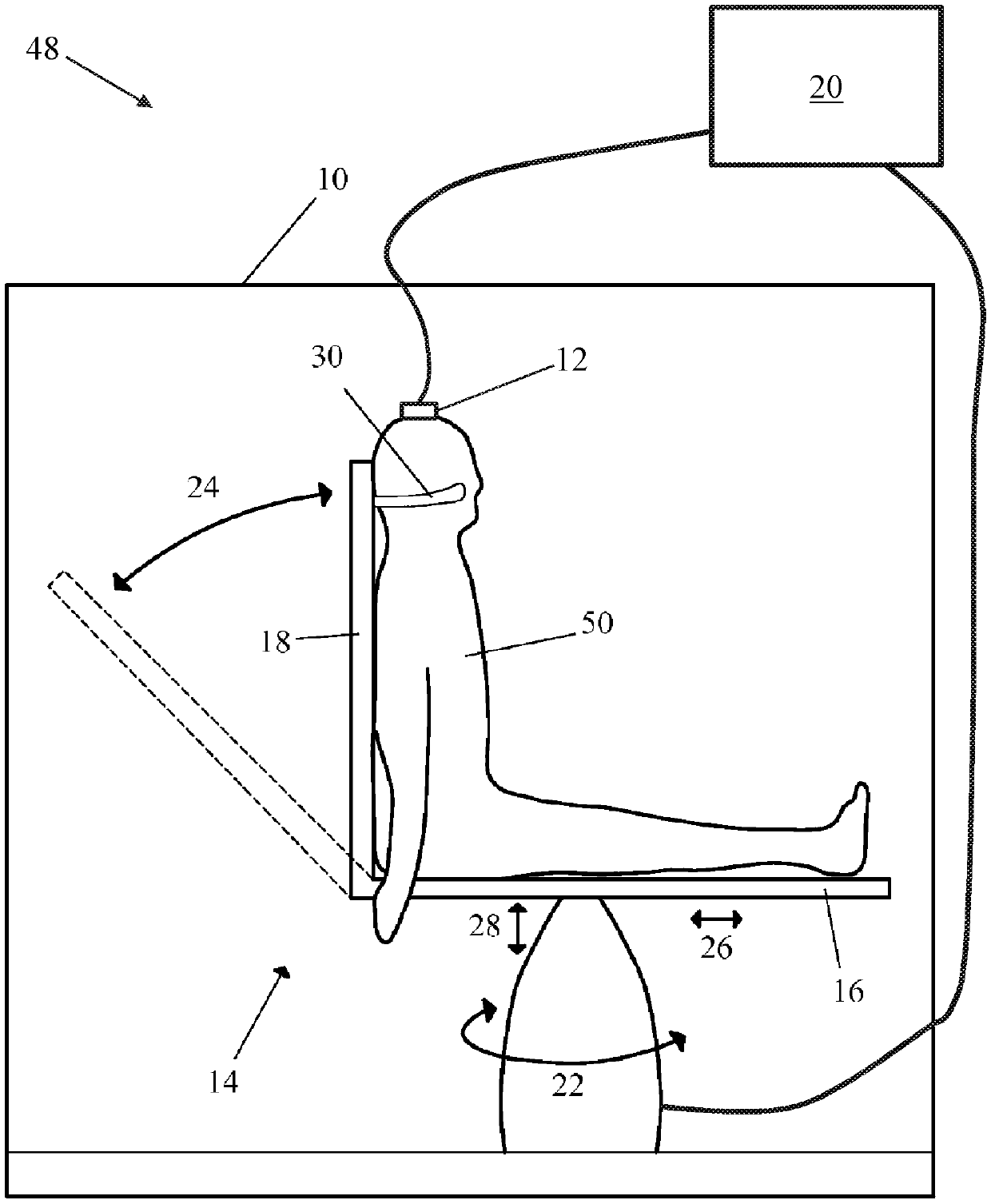
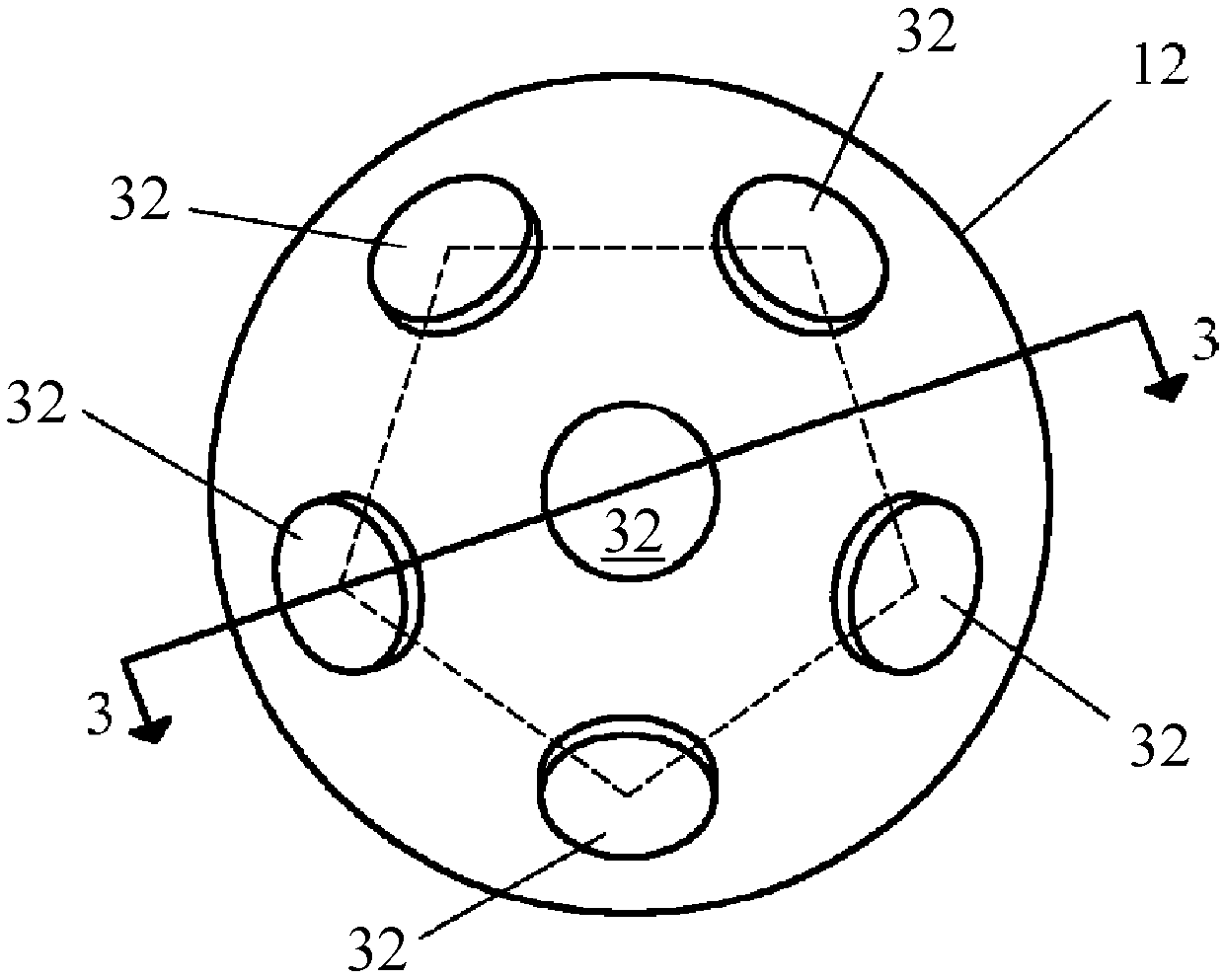
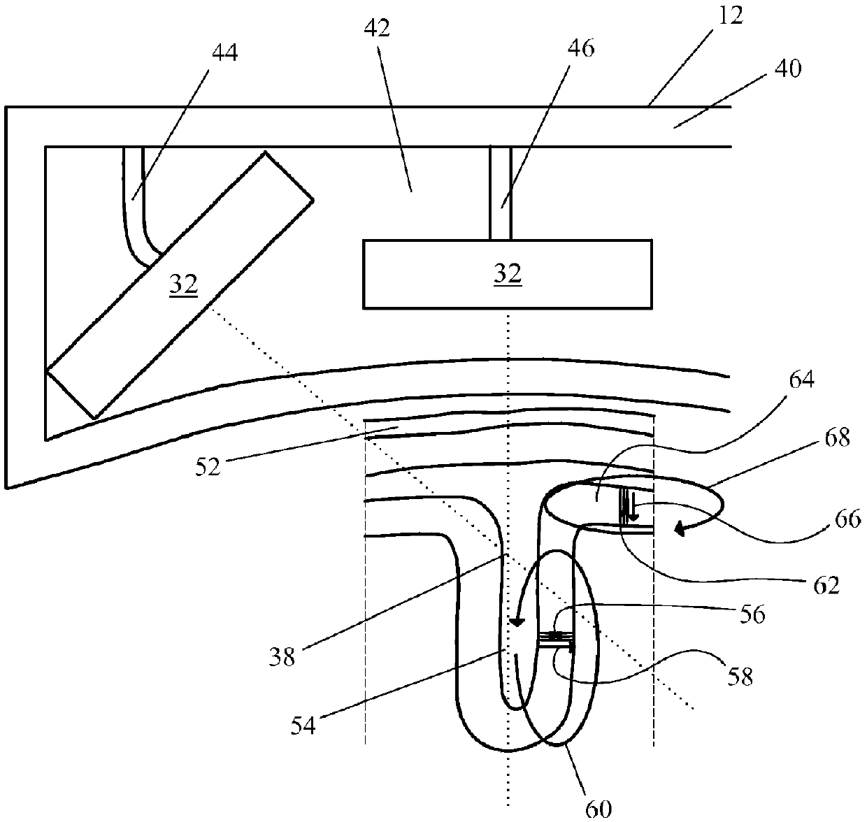
[0044] Figure 1A A magnetoencephalography ("MEG") system 48 for detecting electrical activity in the human brain in the form of a magnetic field generated by the electrical activity is shown, according to one embodiment. The test patient 50 is seated in the patient support device 14 . The Faraday cage 10 surrounds the test patient 50 and the patient support device 14 to block external ambient magnetic fields. sensor head 12 and for cooling the sensor 32 (see Figure 1B ) associated Dewar shell 40 (see Figure 1C ) is fixed in space. The sensor head 12 and patient support device 14 are in communication with and controlled by a computer 20 , which is located outside the Faraday cage 10 .

[0045] Patient support device 14 includes a seat portion 16 and a back portion 18 . The patient support device 14 is rotatable 22 at least a full 360°, wherein the backrest portion 18 is tiltable 24, preferably from a vertical position to a position about 4...


PUM


Abstract

Techniques are described for determining cognitive impairment, an example of which includes accessing a set of epochs of magnetoencephalography (MEG) data of responses of a brain of a test patient toa plurality of auditory stimulus events; processing the set of epochs to identify parameter values one or more of which is based on information from the individual epochs without averaging or otherwise collapsing the epoch data. The parameter values are input into a model that is trained based on the parameters to determine whether the test patient is cognitively impaired.

Description

[0001] Cross References to Related Applications [0002] This application claims U.S. Provisional Application No. 62 / 315,376, filed March 30, 2016, entitled "METHODS AND MAGNETIC IMAGING DEVICES TO INVENTORY HUMAN BRAIN CORTICAL FUNCTION," and filed March 28, 2017, entitled "METHODS AND MAGNETIC IMAGING DEVICES TOINVENTORY HUMAN BRAIN CORTICAL FUNCTION," the priority of U.S. Application No. 15 / 472,130, the entire contents of which are incorporated herein by reference. technical field [0003] This specification relates to the field of medical imaging. More specifically, the present specification relates to systems and methods for detecting and evaluating electrical activity in the brain. Background technique [0004] Despite the rapidly increasing burden on society, progress in developing treatments for neurodegenerative diseases, such as Alzheimer's disease ("AD"), has been slow. [0005] Part of the challenge in developing effective therapeutics is the requirement that ...

Claims


Application Information

Patent Timeline
Patent Type & Authority Applications(China)
IPC IPC(8): A61B5/055A61B5/00A61B5/04A61B5/0484
CPCA61B5/7203A61B5/7264A61B5/0042A61B5/055A61B5/162A61B5/6803A61B2503/40A61B2560/0223A61B2562/0223A61B2576/026A61B5/7257A61B5/4088A61B5/7267A61P25/28G16H30/40G16H50/20G16H50/70A61B5/246
Inventor J·P·福德G·P·萨德雷
Owner BRAIN F I T IMAGING LLC
Why Eureka
Try Eureka