A Semi-Automatic Brain Image Segmentation Method

An image segmentation and semi-automatic technology, applied in image analysis, image enhancement, image data processing, etc., can solve the problem of uneven tissue contour, achieve a wide range of applications, avoid the influence of subjective factors, and reduce the effect of error

Inactive Publication Date: 2019-11-05
HARBIN UNIV OF SCI & TECH
View PDF2 Cites 0 Cited by
  • Summary
  • Abstract
  • Description
  • Claims
  • Application Information

AI Technical Summary

Problems solved by technology

The atlas-based segmentation method can effectively use the prior information of the atlas, so it is widely used in the field of automatic or semi-automatic image segmentation, but the tissue contour obtained by this method is not smooth

Method used


Image

  • A Semi-Automatic Brain Image Segmentation Method
  • A Semi-Automatic Brain Image Segmentation Method
  • A Semi-Automatic Brain Image Segmentation Method

Examples


Embodiment Construction

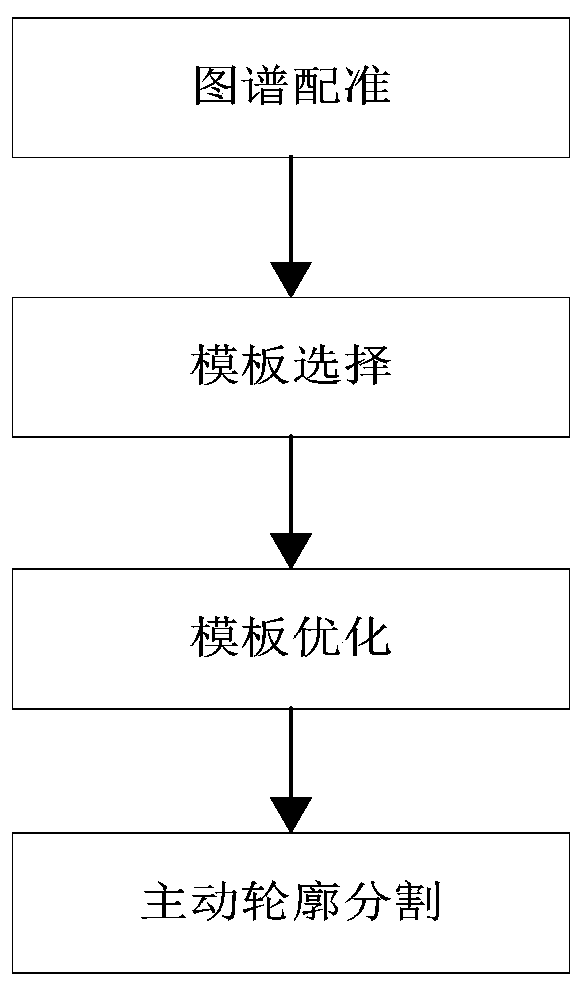
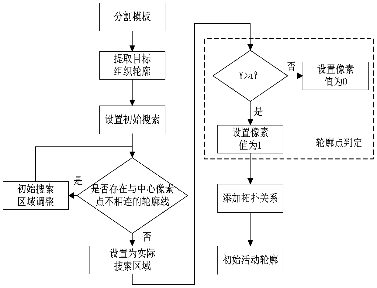
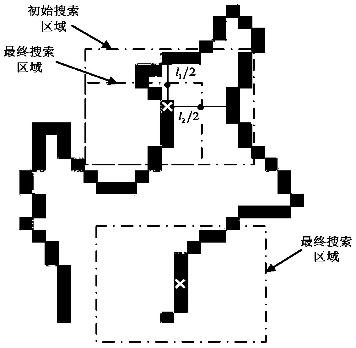
[0054] Such as figure 1 As shown, the specific implementation process of a kind of semi-automatic brain image segmentation method described in this embodiment is:

[0055] Step 1. Atlas registration:

[0056] Given the atlas of the target tissue, the atlas contains N grayscale images F i (i=1,2...N) and the atlas label image L corresponding to the atlas grayscale image i (i=1,2...N), the atlas label image L i for manual grayscale images from the atlas F i The image of the target tissue is marked in the target tissue, and then the target image T and the grayscale image F of each map are combined using an affine transformation-based registration method i Perform registration to obtain the grayscale image F of each map i deformation field;

[0057] Step 2. Template selection:

[0058] Spectrum grayscale image F after measuring deformation i ' and the target image T, select the spectral grayscale image F with the largest similarity value m (m is the label of the spectral ...


PUM


Abstract

The invention discloses a semi-automatic brain image segmentation method, in particular comprising the following steps: firstly, a map registration and a template selection method are used to obtain shape prior information of a target tissue and generate a segmentation template; Secondly, the template optimization method is used to reduce the error in the process of map registration and generate the initial active contour. Finally, the active contour model is used to segment the target tissue. The invention combines the advantages of the atlas registration method and the active contour segmentation method, and realizes the semi-automatic segmentation of the brain image. The method of the invention effectively utilizes shape prior information of the atlas, and can obtain smooth and continuous target tissue outline.

Description

technical field [0001] The invention relates to the technical field of image segmentation, in particular to a semi-automatic brain tissue segmentation method based on MR images. Background technique [0002] Brain image segmentation is crucial to the diagnosis and treatment of brain tissue diseases, and brain image segmentation technology is also the basis for three-dimensional reconstruction of the brain and quantitative analysis of lesions. The accuracy of image segmentation directly affects the location of lesion tissue, the measurement of the shape and size of lesion tissue, and the formulation of clinical diagnosis and treatment plan for brain tissue diseases. [0003] The image segmentation method based on the active contour algorithm has simple expressions, high computational efficiency, and can obtain smooth and continuous target tissue contours. In recent decades, the active contour algorithm has been widely used in image edge detection, medical image segmentation ...

Claims


Application Information

Patent Timeline
Patent Type & Authority Patents(China)
IPC IPC(8): G06T7/149G06T7/00G06T7/38
CPCG06T7/0012G06T7/149G06T7/38G06T2207/30016
Inventor 王沫楠李鹏程
Owner HARBIN UNIV OF SCI & TECH
Why Eureka
Try Eureka