Multifunctional aural rigid endoscope

An endoscopic and multi-functional technology, applied in the field of ear inspection equipment, can solve the problems of high brightness, heating, replacement, and adjustment at any time

Active Publication Date: 2019-02-19
THE FIRST AFFILIATED HOSPITAL OF ZHENGZHOU UNIV
View PDF10 Cites 2 Cited by
  • Summary
  • Abstract
  • Description
  • Claims
  • Application Information

AI Technical Summary

Problems solved by technology

[0004] 1. Observation angle is limited;
[0005] 2. Due to the fine structure of the ear, it is impossible to visually magnify the lesion for diagnosis and treatment;
[0006] 3. The observed situation cannot be imaged and photographed in the form of digital images
[0008] 1. Fogging: Since the rigid endoscope is made of metal, the surface temperature decreases after being soaked in enzyme solution and disinfectant solution, and the human body has body temperature. When the rigid endoscope enters the ear canal, the hot water vapor in the ear canal will liquefy when it is cooled The small water droplets condense on the surface of the lens and look like white mist under the refraction of light, which affects diagnosis and treatment. The traditional solutions are: ①Take out the endoscope, wipe it with iodophor or alcohol cotton ball, and then enter the ear again However, when re-entering, after a short period of use, a fog layer will still be formed; ②Before use, soak the endoscope in physiological saline at about 30 degrees (the soaking solution cannot be reused) for a few minutes, and make its surface temperature and When the body temperature reaches a certain level, enter the human body for observation, but it needs to be heated in time and replace the normal saline, which makes the diagnosis and treatment cumbersome; ③ using anti-fogging agent, but it is expensive and the cost is high
[0009] 2. The condition of the ear canal of different patients is different (especially postoperative patients), resulting in different refraction imaging effects of light, which directly affects the quality of imaging. If the brightness is too low, the image color is dull and unclear; if the brightness is too high, it will cause image overexposed
However, the adjustment key of the light source of the existing rigid endoscope is on the main unit, and the operator cannot adjust it at any time during the operation. cause great inconvenience

Method used


Image

  • Multifunctional aural rigid endoscope
  • Multifunctional aural rigid endoscope
  • Multifunctional aural rigid endoscope

Examples


Embodiment Construction

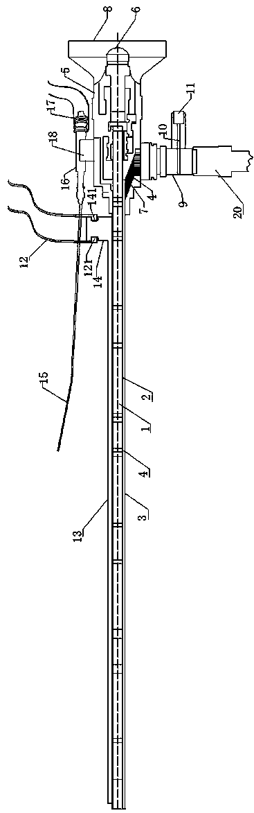
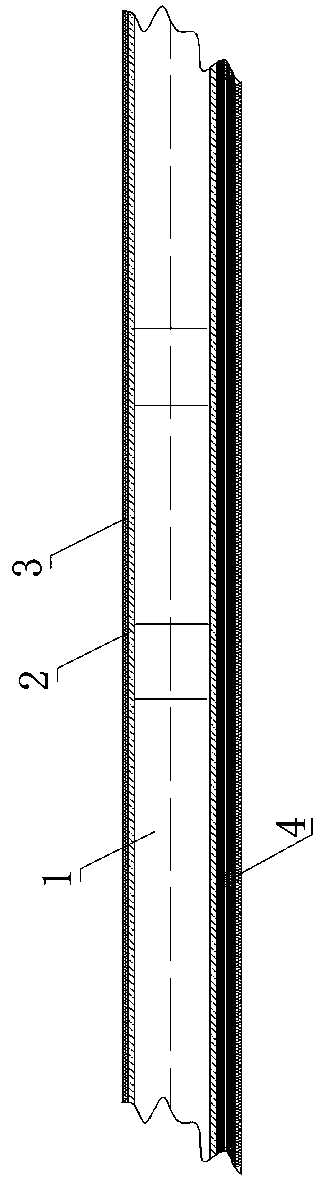
[0027] like figure 1 and figure 2 As shown, the present invention includes an objective lens part, an eyepiece part, an imaging adapter joint 8, an illumination adjustment connection part, a negative pressure defogging part and a negative pressure adsorption needle body. The objective lens part includes the objective lens 1, which is sleeved on the objective lens The inner mirror tube 2 outside 1 and the outer mirror tube 3 outside the inner mirror tube 2 are sleeved in the gap, the front end of the outer mirror tube 3 is a camera head, and the gap between the inner mirror tube 2 and the outer mirror tube 3 forms a wire cavity, A light guide fiber 4 is arranged in the line cavity; the line cavity is located at the lower part of the outer mirror tube 3 , and the right end of the line cavity communicates with the upper end of the line slot 7 .

[0028] The eyepiece portion includes an outer casing 5 and an eyepiece 6 arranged in the outer casing 5. The rear end of the outer mi...


PUM


Abstract

The invention discloses a multifunctional aural rigid endoscope. The multifunctional aural rigid endoscope comprises an objective lens portion, an eyepiece portion, an imaging fitting joint, a lighting adjustment connecting portion, a negative pressure demisting portion and a negative pressure adherence needle. The objective lens portion, the eyepiece portion and a camera mechanism are used for photographing. During photographing, by the negative pressure demisting portion, water vapor in a human body can be sucked into a negative pressure demisting tube, and then discharged by a first rubberhose at the rear end of the negative pressure demisting tube into a collection bottle to achieve demisting, wherein the front end of a horizontal sucker of the negative pressure demisting portion is arranged beside the front end of an outer endoscope tube. During image photographing, a light adjustment piece is adjusted through a knob, the light adjustment piece and the knob are in meshed connection, and the light adjustment piece is rotated slightly while the knob is rotated. The negative pressure adherence needle is used for adhering and cleaning dirt in the ear; when the operation is completed, the medical staff put a needle body into a needle clamping slot through a clamping portion to prevent accidental injury to others.

Description

technical field [0001] The invention relates to the technical field of ear inspection equipment, in particular to a multifunctional ear rigid endoscope. Background technique [0002] At present, forehead mirrors, electric otoscopes and rigid endoscopes are widely used in clinical otology for inspection. [0003] Frontal mirrors and electric otoscopes have the following deficiencies: [0004] 1. Observation angle is limited; [0005] 2. Due to the fine structure of the ear, it is impossible to visually enlarge the lesion for diagnosis and treatment; [0006] 3. The observed situation cannot be imaged and photographed in the form of a digital image. [0007] The rigid endoscope in the prior art includes an endoscope tube, an outside mirror tube, an imaging system and an illumination system. The inner mirror tube is arranged in the outer mirror tube. The outer mirror tube connects the imaging system and the lighting system, and is the part inserted into the ear canal. Use...

Claims


Application Information

Patent Timeline
Patent Type & Authority Applications(China)
IPC IPC(8): A61B1/227A61B1/04A61B1/06A61F11/00
CPCA61B1/00064A61B1/00131A61B1/00165A61B1/00195A61B1/04A61B1/0661A61B1/227A61F11/006
Inventor 郭晨辉叶放蕾叶琳向明丽
Owner THE FIRST AFFILIATED HOSPITAL OF ZHENGZHOU UNIV
Why Eureka
Try Eureka