Disposable oocyte collector
A disposable technology for oocytes, applied in the field of medical devices, can solve the problems of unclear ultrasound imaging, real-time observation of needle insertion depth, and reduced effectiveness, and achieve good surface finish, good puncture feel, and reduced puncture resistance. Effect
- Summary
- Abstract
- Description
- Claims
- Application Information
AI Technical Summary
Problems solved by technology
Method used
Image

Examples
Embodiment 1
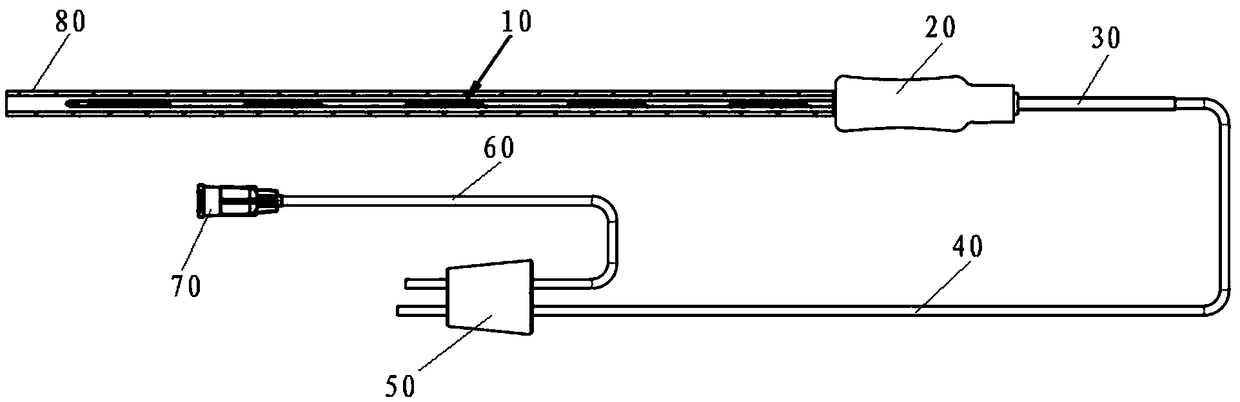
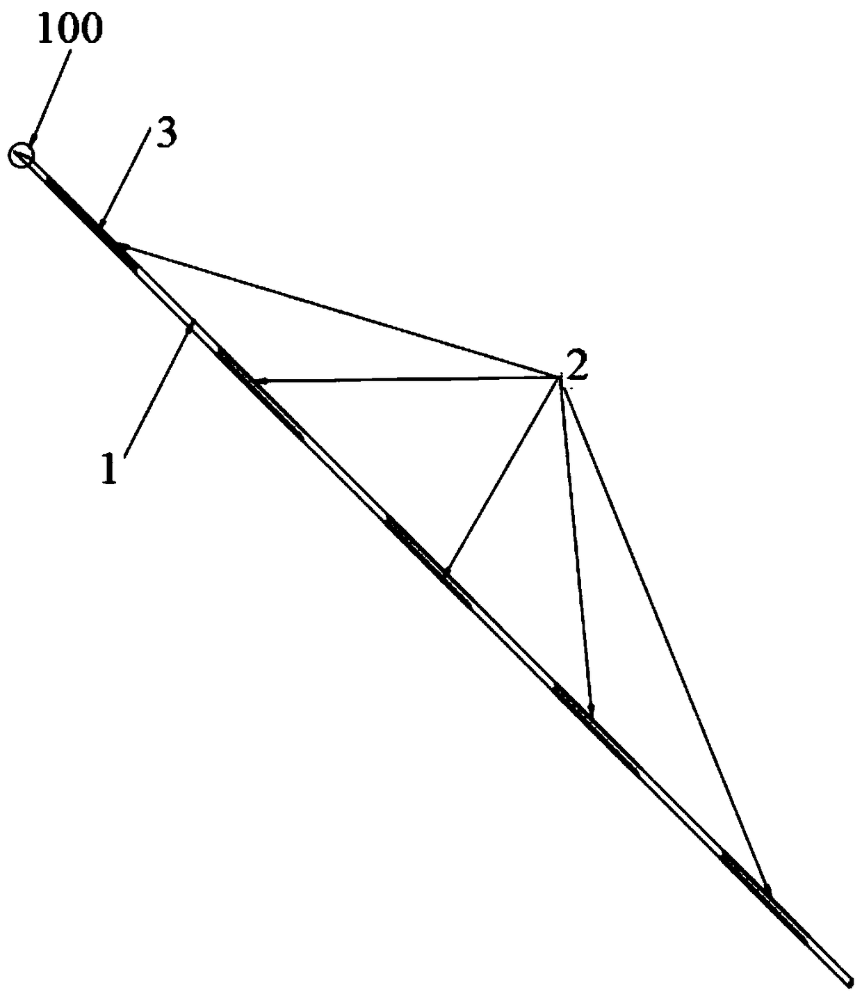
[0044] see Figure 1 to Figure 6 , a disposable oocyte collector, comprising a puncture needle 10, a handle 20, a catheter sleeve 30, a suction catheter 40 and a silicone plug 50 connected in sequence, the silicone plug 50 is connected to a conical joint 70 through a vacuum catheter 60, and the puncture needle 10 is sleeved with a protective sheath 80. The puncture needle 10 includes a needle tube 1 and a needle point 100 connected thereto. An ultrasound imaging enhancement band group 2 is arranged on the outer wall of the needle tube 1; Enhancement bands 3, several ultrasonic imaging enhancement bands 3 are distributed on the outer wall of the needle tube 1 at equal intervals along the axial direction of the needle tube 1; it is known that the length of the puncture needle used to collect oocytes in egg retrieval is mostly 30 cm It can be determined according to the requirements of use. The length of the puncture needle in this embodiment is preferably 30 cm.
[0045] see a...
Embodiment 2
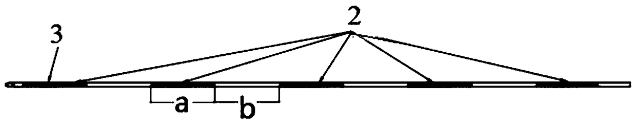
[0050] see Figure 7 , a disposable oocyte collector, which is roughly the same as the disposable oocyte collector in Embodiment 1, except that the length of the puncture needle is 35 cm. The axial length a' of the ultrasonic development enhancement band 3 along the needle tube 1 is 40 mm, and the distance b' between every two adjacent ultrasonic development enhancement bands is 30 mm. The cross-sectional shape of the linear groove 43 is a semicircle, that is, the shape of the linear groove 43 on the cross section of the needle tube 1 is a semicircle. The linear groove 43 has a width of 90um and a depth of 15um.
[0051] The linear groove group 4 is processed by a femtosecond laser with a pulse width of 8ps; the diameter of the femtosecond laser spot during processing is 10um, the wavelength is 1028nm, the output power does not exceed 8W, and the repetition frequency is 10KHz-1000KHz. The laser scanning speed is 0.1~1mm / s.
Embodiment 3
[0053] see Figure 8a and Figure 8b , a single-use oocyte collector, on the basis of Embodiment 1, also includes a flushing catheter 90 and a locking joint 91, and the locking joint 91 is connected to the handle 20 through the flushing catheter 90; the puncture needle 10 includes The inner needle 12 and the outer needle 11 are arranged coaxially to the outside, the suction catheter 10 communicates with the inner needle 12 through the catheter sleeve 30 and the handle 30 in turn, and the irrigation catheter 90 passes through the handle 20 and the channel between the inner needle 12 and the outer needle 11 13 communicates; the outer needle 11 includes a needle tube 1 and a needle tip 100, so that the ultrasonic imaging enhancement band group 2 is arranged on the outer wall of the needle tube 1 of the outer needle 11.
[0054] The disposable oocyte collector of the present invention adopts a linear groove 43 with a semicircular section to form a linear groove group 4, and divid...
PUM
| Property | Measurement | Unit |
|---|---|---|
| Length | aaaaa | aaaaa |
| Width | aaaaa | aaaaa |
| Depth | aaaaa | aaaaa |
Abstract
Description
Claims
Application Information
 - Generate Ideas
- Intellectual Property
- Life Sciences
- Materials
- Tech Scout
- Unparalleled Data Quality
- Higher Quality Content
- 60% Fewer Hallucinations
Browse by: Latest US Patents, China's latest patents, Technical Efficacy Thesaurus, Application Domain, Technology Topic, Popular Technical Reports.
© 2025 PatSnap. All rights reserved.Legal|Privacy policy|Modern Slavery Act Transparency Statement|Sitemap|About US| Contact US: help@patsnap.com