Thymic cell image segmentation method based on improved U-Net network

A technology for thymocyte and image segmentation, which is applied in the field of image processing, can solve the problem of low thymus image and achieve the effect of excellent robustness and fast segmentation speed

Inactive Publication Date: 2019-01-11
HANGZHOU DIANZI UNIV
View PDF6 Cites 27 Cited by
  • Summary
  • Abstract
  • Description
  • Claims
  • Application Information

AI Technical Summary

Problems solved by technology

[0005] In order to solve the above problems, the present invention creates a new cell image segmentation method by improving a basic segmentation network, w

Method used


Image

  • Thymic cell image segmentation method based on improved U-Net network
  • Thymic cell image segmentation method based on improved U-Net network
  • Thymic cell image segmentation method based on improved U-Net network

Examples


Embodiment Construction

[0045] In order to make the object, technical solution and advantages of the present invention clearer, the present invention will be further described in detail below in conjunction with the accompanying drawings and embodiments. It should be understood that the specific embodiments described here are only used to explain the present invention, not to limit the present invention.

[0046] On the contrary, the invention covers any alternatives, modifications, equivalent methods and schemes made within the spirit and scope of the invention as defined by the claims. Further, in order to make the public have a better understanding of the present invention, some specific details are described in detail in the detailed description of the present invention below. The present invention can be fully understood by those skilled in the art without the description of these detailed parts.

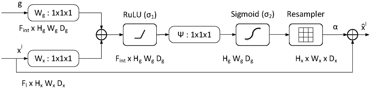
[0047] see figure 1 , figure 1 The technical scheme of the present invention is based on the st...


PUM


Abstract

The present invention discloses a thymic cell image segmentation method based on an improved U-Net network, which comprises the following steps: carrying out image preprocessing on a UCSB breast imagedata set; in a U-Net network, adding a void residual module and an attention module; training the U-NET network according to the set training strategy; setting up the evaluation indexes including F1score, object-level Dice coefficient and Hausdorff distance. The optimal model is obtained by optimizing the network through the evaluation indexes. The cell image to be segmented is inputted into theoptimal model, and the segmentation mask is obtained by feature extraction and feature upsampling. By improving a basic segmentation network and creating a new cell image segmentation method, the invention solves the problem of low precision in the automatic segmentation process of the thymus image, and improves the accuracy and efficiency of the segmentation.

Description

technical field [0001] The invention belongs to the field of image processing and relates to a thymocyte image segmentation method based on an improved U-Net network. Background technique [0002] In recent years, the incidence of colon cancer has increased. Colorectal cancer is the third most common cancer in men and the second most common cancer in women. Approximately 95% of colorectal cancers are adenocarcinomas. Typically, a typical gland consists of a luminal region forming an inner tubular structure and an epithelial nucleus surrounding the cytoplasm. Malignancies arising from the glandular epithelium, also known as adenocarcinomas, are the most prevalent form of cancer. On histopathological examination, glandular morphology is widely used to evaluate several adenocarcinomas, including breast, prostate, and colon. Accurate gland segmentation is a key prerequisite for obtaining reliable morphological statistics that indicate tumor aggressiveness. Previously, gland s...

Claims


Application Information

Patent Timeline
IPC IPC(8): G06T7/11
CPCG06T2207/20081G06T2207/20084G06T2207/30004G06T7/11
Inventor 于海滨贝琛圆潘勉吕帅帅和文杰于彦贞刘爱林李子璇
Owner HANGZHOU DIANZI UNIV
Why Eureka
Try Eureka