
A quantum dot-based biomolecule concentration detection method

A technology for biomolecules and concentration detection, applied in the direction of measuring devices, analytical materials, material excitation analysis, etc., can solve the problems that the instrument has not been marketed, it is difficult to find quantum dots, quantum dot fluorescence intensity and wavelength inhomogeneity

Active Publication Date: 2020-10-27
傅英
View PDF17 Cites 0 Cited by
  • Summary
  • Abstract
  • Description
  • Claims
  • Application Information

AI Technical Summary

Problems solved by technology

And in practical optical measurement applications, quantitative measurement of small changes in fluorescence intensity and fluorescence wavelength is difficult to achieve
At the same time, the synthesis of quantum dots is affected by many experimental conditions. Even if the same batch of quantum dots is grown, it is difficult to find quantum dots with the same fluorescence intensity and wavelength. Therefore, the quantum dots used for biomolecular inspection have different fluorescence intensity and wavelength. Uniformity, which brings insurmountable difficulties to the realization of instruments that use the changes in the fluorescence intensity and wavelength of quantum dots to detect biomolecules
Therefore, the patent application for quantum dot biological detection instruments based on this method has been proposed since 2000, but the marketization of this type of instrument has not been realized until now. Fluorescence intensity and wavelength changes after molecules are difficult to quantitatively identify QD-Ab-biomolecules

Method used


Image

  • A quantum dot-based biomolecule concentration detection method
  • A quantum dot-based biomolecule concentration detection method
  • A quantum dot-based biomolecule concentration detection method

Examples


Embodiment 2

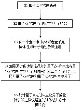
[0049] The method for detecting the concentration of biomolecules based on quantum dots involved in this embodiment includes the following steps:

[0050] S1. According to the water-soluble quantum dot QD surface modification (carboxyl, sulfhydryl), through different cross-linking agents and the antibody coupling of the target biomolecule to be detected:

[0051] Using the SMCC (succinimidyl 4-[N-maleimidomethyl]cycloethane-1-carboxylate Eurolink reaction to couple sulfhydryl-activated quantum dots to amino-activated antibodies for amino and sulfhydryl The covalent reaction couples the antibody and the quantum dots to form a quantum dot-antibody stock solution.

[0052] S2. Stir the quantum dot-antibody stock solution with the solution to be tested containing the target biomolecule for 20 minutes, mix and culture to combine the quantum dot-antibody with the target biomolecule, and obtain a mixture containing quantum dot-antibody and quantum dot-antibody-biomolecule solution; ...

Embodiment 3

[0059] The method for detecting the concentration of biomolecules based on quantum dots involved in this embodiment includes the following steps:

[0060] S1. According to the water-soluble quantum dot QD surface modification (carboxyl, sulfhydryl), through different cross-linking agents and the antibody coupling of the target biomolecule to be detected:

[0061] Using the SMCC (succinimidyl 4-[N-maleimidomethyl]cycloethane-1-carboxylate Eurolink reaction to couple sulfhydryl-activated quantum dots to amino-activated antibodies for amino and sulfhydryl The covalent reaction couples the antibody and the quantum dots to form a quantum dot-antibody stock solution.

[0062] S2. Stir the quantum dot-antibody stock solution with the solution to be tested containing the target biomolecule for 20 minutes, mix and culture to combine the quantum dot-antibody with the target biomolecule, and obtain a mixture containing quantum dot-antibody and quantum dot-antibody-biomolecule solution; ...


PUM


Abstract

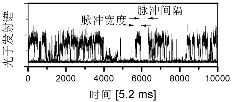
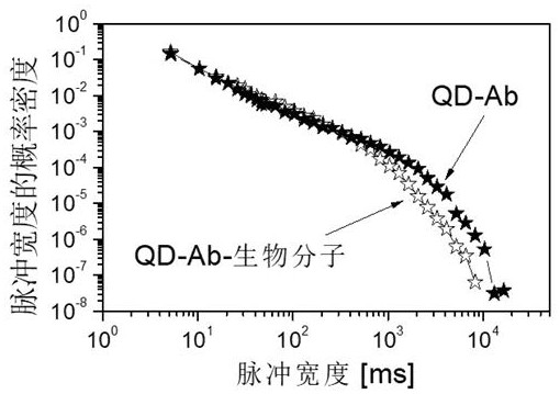
The invention discloses a method of detecting biomolecule concentration based on quantum dots. The method comprises the following steps of: coupling quantum dots with an antibody of a target biomolecule to be detected, combining the quantum dot-antibody with the target biomolecule through the mixed cultivation of a quantum dot-antibody stock solution and a target biomolecule-containing solution tobe detected, enabling a mixed solution to pass through a microfluidic channel, and controlling the flow rate of the mixed solution to cause the quantum dot-antibody or the quantum dot-antibody-biomolecule to pass the microfluidic channel; measuring the time resolution single photon characteristic spectrum of the quantum dot-antibody or the quantum dot-antibody-biomolecule passing the microfluidicchannel, and distinguishing the quantum dot-antibody and the quantum dot-antibody-biomolecule; and calculating the concentration of the target biomolecule in the solution to be detected. The method can detect biomolecules of fM concentration, and the detection speed is greatly increased.

Description

technical field [0001] The invention relates to a method for detecting the concentration of biomolecules, in particular to a method for detecting the concentration of biomolecules based on quantum dots. Background technique [0002] The rapid development of society has greatly improved people's living standards, and put forward new requirements for living hygiene and disease prediction. However, it is difficult for existing biomolecular detection methods to achieve rapid identification and measurement. Therefore, in the monitoring of bacteria such as drinking water, It is difficult to make breakthroughs in practical applications in the screening of bacterial drug resistance and the identification and quantification of cancer biomarkers. State-of-the-art methods for identifying and quantifying biomolecules at low concentrations include: [0003] Liquid chromatography and mass spectrometry (LC-MS): LC physically separates components in a liquid solution; MS provides structura...

Claims


Application Information

Patent Timeline
Patent Type & Authority Patents(China)
IPC IPC(8): G01N21/64
CPCG01N21/6402
Inventor 傅英杨希峰
Owner 傅英
Learn More