Fixing method of pathological specimen

A fixation method and specimen technology, applied in the field of pathological specimen processing, can solve the problems of affecting the exposure of antigenic determinants, hindering the length of nucleic acid extraction, and affecting the diagnosis results, and achieve the effects of good fixation effect, shortened fixation time, and low use cost.

Active Publication Date: 2018-11-06
LUOHE MEDICAL COLLEGE
View PDF10 Cites 3 Cited by
  • Summary
  • Abstract
  • Description
  • Claims
  • Application Information

AI Technical Summary

Problems solved by technology

However, the use of formaldehyde to fix tissues mainly has the following disadvantages: 1. It is very volatile, and its volatility is accelerated with the increase of ambient temperature. Formaldehyde ranks second in the current list of toxic chemicals in my country, and has been identified as a Carcinogens and teratogens, especially doctors and technicians working on the front line, are exposed to formaldehyde seriously exceeding the standard for a long time, causing serious harm to health
②It is easy to cause extensive protein cross-linking in the tissue, which affects the exposure of antigenic determinants in immunohistochemical staining
③It is easy to cause DNA breakage, cross-linking hinders the extraction of nucleic acids and limits the length of PCR amplification products of DNA fragments
However, this fixative solution contains a large amount of ethanol, which has a syneresis effect on the specimen. Long-term use will easily cause the specimen to lose water, and the shape of the specimen after fixation will change greatly, sometimes affecting the diagnostic results.

Method used


Image

  • Fixing method of pathological specimen
  • Fixing method of pathological specimen
  • Fixing method of pathological specimen

Examples


Embodiment 1~ Embodiment 3

[0028] Embodiment 1~Example 3: Take the pathological tissue taken from human thyroid (1-1.5cm)×1cm×(0.2-0.3cm) as an example.

[0029] A method for fixing a pathological specimen, comprising the following steps:

[0030] S1: After cleaning the fixed cylinder, clean it with carbolic water with a mass fraction of 6%~8%, and dry it at room temperature;

[0031] S2: prepare fixative;

[0032]S3: Add the fixative solution obtained in step S2 to the fixation cylinder in step S1, put the fixation cylinder in a constant temperature box, and store at a constant temperature of 42°C for 2 hours. The volume of the fixation solution is 6 to 10 times the volume of the specimen to ensure Can be completely immersed in the fixative;

[0033] S4: Put the pathological specimen in the fixative solution for 1.5 hours, and then take it out.

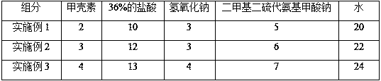
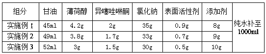
[0034] The compositions of Examples 1-3 fixatives are shown in Table 1 and Table 2 below.

[0035] Table 1 Components and weights in the fixative

[0036...

Embodiment 4~ Embodiment 6

[0042] Embodiment 4~Example 6: Take the pathological tissue taken from human liver (1-1.5cm)×1cm×(0.2-0.3cm) as an example.

[0043] A method for fixing a pathological specimen, comprising the following steps:

[0044] S1: After cleaning the fixed cylinder, clean it with carbolic water with a mass fraction of 6%~8%, and dry it at room temperature;

[0045] S2: prepare fixative;

[0046] S3: Add the fixative solution obtained in step S2 into the fixation cylinder in step S1, put the fixation cylinder in a constant temperature box, store at a constant temperature of 48°C for 1.5h, the volume of the fixation solution is 6-10 times the volume of the specimen, ensure The specimen can be completely immersed in the fixative solution;

[0047] S4: Put the pathological specimen in the fixative solution for 1 hour and take it out.

[0048] Wherein, the composition of embodiment 4~6 stationary solution is as shown in table 3 below.

[0049] Table 3 Components and weights in the fixat...

Embodiment 7~ Embodiment 9

[0054] Embodiment 7 to Embodiment 9: Take the pathological tissue taken from (1-1.5cm)×1cm×(0.2-0.3cm) human breast as an example.

[0055] A method for fixing a pathological specimen, comprising the following steps:

[0056] S1: After cleaning the fixed cylinder, clean it with carbolic water with a mass fraction of 6%~8%, and dry it at room temperature;

[0057] S2: prepare fixative;

[0058] S3: Add the fixative solution obtained in step S2 into the fixation cylinder in step S1, put the fixation cylinder in a constant temperature box, and store at a constant temperature of 45°C for 1.5h. The volume of the fixation solution is 6 to 10 times the volume of the specimen to ensure The specimen can be completely immersed in the fixative solution;

[0059] S4: Put the pathological specimen in the fixative solution for 1.5 hours, and then take it out.

[0060] Wherein, the composition of the stationary solution of Example 7 is the same as that of Example 4, the composition of the...


PUM

PropertyMeasurementUnit
diameteraaaaaaaaaa

Abstract

The invention discloses a fixing method of a pathological specimen and belongs to the technical field of pathological specimen treatment. The fixing method comprises the following steps: after a fixedcylinder is cleaned, cleaning with 6-8 percent by weight of phenolated water and air-drying at a normal temperature; preparing a fixing solution; adding the fixing solution in the fixed cylinder, putting the fixed cylinder in a constant-temperature cabinet and preserving for 1.5-2 hours at a constant temperature of 42-18 DEG C; putting the pathological specimen in the fixing solution to fix for 1-1.5 hours and then taking out. The fixing solution prepared by the fixing method disclosed by the invention does not contain formaldehyde, is stable in performance, environmentally-friendly, safe andgood in fixing effect and has a significant inhibiting effect on common bacteria. The fixing method disclosed by the invention can well preserve physiological and pathological morphological structures as well as biochemical and immunohistochemical components of tissue cells in vitro, is short in fixing time, sufficient in fixing, lays a solid foundation for making excellent pathological sectionsand is also a basis for the success of technical methods such as special staining, histochemistry, immunohistochemistry and tissue in-situ molecular hybridization.

Description

technical field [0001] The invention belongs to the technical field of pathological specimen processing, and in particular relates to a method for fixing pathological specimens. Background technique [0002] Pathological specimens are important materials for morphological teaching and scientific research, as well as an important basis for tumor diagnosis, and tissue fixation is the key to making diagnostic slices. The pathological diagnosis of living tissue is the gold standard for the diagnosis of surgical diseases. If the tissue is autolyzed, the structure is destroyed, and the cells are deformed, the diagnosis may not be possible. Pathological specimens should be properly fixed in time after isolation, and then sent to the pathology department for examination. Improper handling of pathological specimens will not only bring difficulties in clinical diagnosis, but also affect the treatment effect of patients. [0003] At present, the pathology departments of most hospitals...

Claims


Application Information

Patent Timeline
Patent Type & Authority Applications(China)
IPC IPC(8): G01N1/30
CPCG01N1/30G01N2001/307
Inventor 刘春灵程琦申现锋
Owner LUOHE MEDICAL COLLEGE
Why Eureka
Try Eureka