Detection method of protein marker for urine exosome

A protein labeling and detection method technology, applied in the field of medical testing, can solve the problems of poor diagnostic significance and lack of specificity

Pending Publication Date: 2018-10-02
THE SECOND AFFILIATED HOSPITAL OF NANJING MEDICAL UNIV
View PDF5 Cites 2 Cited by
  • Summary
  • Abstract
  • Description
  • Claims
  • Application Information

AI Technical Summary

Problems solved by technology

Although most of the urine exosomes come from nephrons, their source cells are still diverse, and the analysis of urine exosome changes without distinction lacks specificity, so the diagnostic significance of the disease is poor

Method used


Image

  • Detection method of protein marker for urine exosome
  • Detection method of protein marker for urine exosome

Examples


Embodiment Construction

[0028] In order to make the object, technical solution and advantages of the present invention clearer, the present invention will be further described in detail below in conjunction with the accompanying drawings and embodiments. It should be understood that the specific embodiments described here are only used to explain the present invention, not to limit the present invention.

[0029] A method for detecting protein markers of urine exosome, the method comprising the following steps:

[0030] (1) Extract and purify the exosome from freshly collected urine, collect the precipitate and resuspend to obtain the exosome suspension;

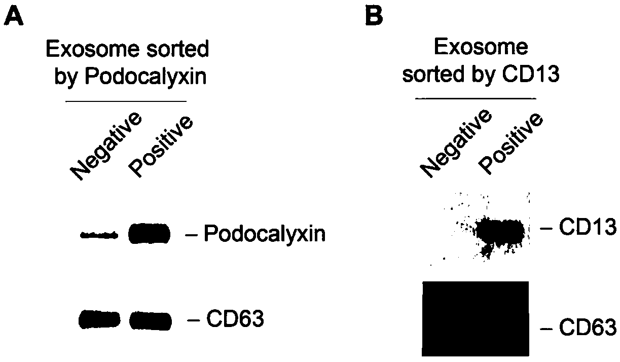
[0031] (2) Separately use CD13 and podocalyxin-coated magnetic beads (purchased from BDPharmingen, USA) to sort the exosome suspension obtained in step (1) to obtain the exosome sorting liquid;

[0032] (3) the sorting liquid that step (2) obtains is passed through with BCA kit ( BCAProtein AssayKit, NCI3225CH) to measure the protein concentrati...


PUM


Abstract

The invention discloses a detection method of a protein marker for urine exosome. The method comprises the following steps: extracting and purifying exosome from freshly collected urine, collecting precipitate, and performing resuspending, so as to obtain exosome suspension; performing separation on the exosome suspension obtained in the step (1) through magnetic beads coated by CD13 and podocalyxin respectively, so as to obtain an exosome separation fluid; measuring the protein concentration of exosome of the separation fluid through a BCA kit; preparing the separation fluid into a protein loading sample according to the protein concentration, and performing CD13, Podocalyxcin and CD63westernblot detection on the sample, so as to obtain the required protein marker after identification. The method has the advantages that positive exosome of podocalyxin and CD13 in urine can be respectively extracted, and the method is beneficial to further analysis and detection to the exosome, so thatchange of sertoli cell and proximal renal tubular epithelial cell can be accurately reflected, and a biological marker is provided for diagnosis and treatment of diseases.

Description

technical field [0001] The invention belongs to the technical field of medical detection, and in particular relates to a method for detecting protein markers of urine exosome. Background technique [0002] In recent years, the incidence of various acute and chronic kidney injuries has been increasing year by year, and nearly 1% of patients are complicated with acute kidney injury during hospitalization, and the number of patients with chronic kidney disease is more than 100 million, which has become an important cause of death for Chinese people . Early diagnosis and targeted treatment are the key to preventing acute kidney injury and delaying the progression of chronic kidney disease. However, the poor sensitivity of traditional renal injury diagnostic methods and the poor diagnostic accuracy of new renal injury biomarkers make it impossible for people to timely and accurately determine whether renal injury occurs and its degree, pathological type, disease progression and ...

Claims


Application Information

Patent Timeline
IPC IPC(8): G01N33/68
CPCG01N33/68
Inventor 杨俊伟周阳曹红娣熊明霞江蕾闻萍方丽何伟春叶红
Owner THE SECOND AFFILIATED HOSPITAL OF NANJING MEDICAL UNIV
Why Eureka
Try Eureka