Multimodality mineralogy segmentation system and method

A multi-modal imaging, mineral technology, applied in the analysis of materials, image analysis, material analysis using wave/particle radiation, etc., can solve the problem of no mineral identification method, etc.

Pending Publication Date: 2018-08-21
CARL ZEISS X RAY MICROSCOPY
View PDF9 Cites 2 Cited by
  • Summary
  • Abstract
  • Description
  • Claims
  • Application Information

AI Technical Summary

Problems solved by technology

Although CT intensity correlates with mineral density, there is no direct method for mineral identification on X-ray CT microscopy systems

Method used


Image

  • Multimodality mineralogy segmentation system and method
  • Multimodality mineralogy segmentation system and method
  • Multimodality mineralogy segmentation system and method

Examples


Embodiment Construction

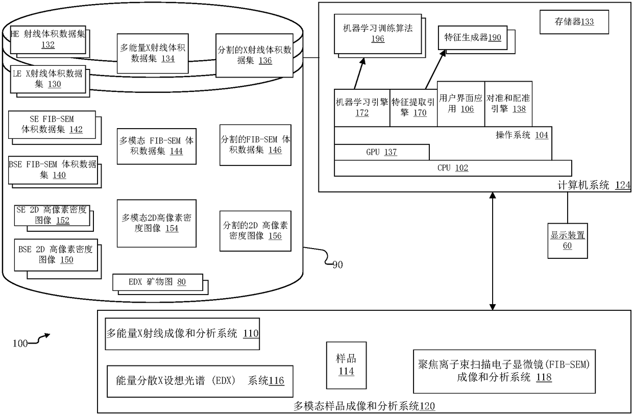
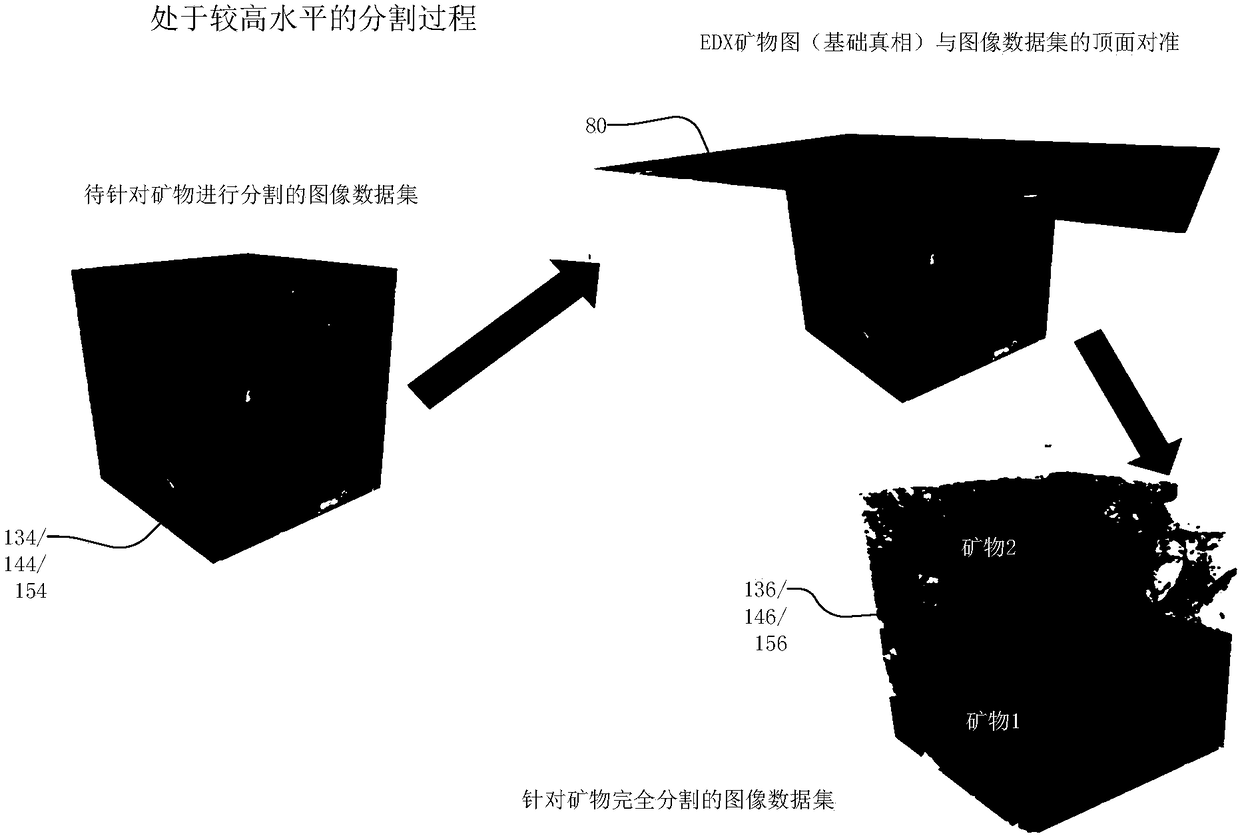
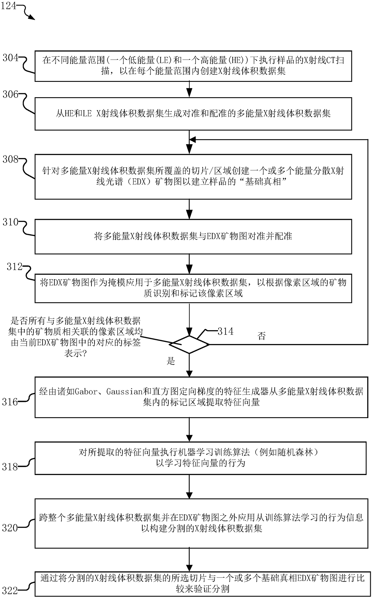
[0030] The invention will be described more fully hereinafter with reference to the accompanying drawings, in which illustrative embodiments of the invention are shown. However, this invention may be embodied in many different forms and should not be construed as limited to the embodiments set forth herein; rather, these embodiments are provided so that this disclosure will be thorough and complete, and will fully convey to those skilled in the art scope of the invention.

[0031] As used herein, the term "and / or" includes any and all combinations of one or more of the associated listed items. Furthermore, unless expressly stated otherwise, the singular forms and the articles "a" and "the" are intended to include the plural forms as well. It should also be understood that the terms comprise and / or comprise, when used in this specification, indicate the presence of stated features, integers, steps, operations, elements and / or elements, but do not exclude one or more other feat...


PUM


Abstract

A multimodality imaging system and method for mineralogy segmentation is disclosed. Image datasets of the sample are generated for one or more modalities, including x-ray and focused ion beam scanningelectron microscope (FIB-SEM) modalities. Mineral maps are then created using Energy Dispersive X-ray spectroscopy (EDX) from at least part of the sample covered by the image datasets. The EDX mineral maps are applied as a mask to the image datasets to identify and label regions of minerals within the sample. Feature vectors are then extracted from the labeled regions via feature generators suchas Gabor filters. Finally, machine learning training and classification algorithms such as Random Forest are applied to the extracted feature vectors to construct a segmented image representation of the sample that classifies the minerals within the sample.

Description

Background technique [0001] Various imaging modalities in two dimensions (2D) and three dimensions (3D) have been used to identify and visualize rock mineral content. For example, these imaging modalities can analyze rock samples from oil and gas extraction operations to determine porosity and minerals, thereby simulating the flow and mechanical properties of samples obtained during exploration and production operations. Generally, these imaging modalities are characterized by destructive and non-destructive techniques. Furthermore, some modalities only analyze surface features, while others can analyze three-dimensional structures. [0002] In typical operation, these imaging modalities create image datasets such as 3D volumes or 2D images. Image analysis techniques are then employed to infer mineral content from the volumes and images created by the different imaging modalities. [0003] Non-destructive imaging systems include X-ray computed tomography (CT) microscopes an...

Claims


Application Information

Patent Timeline
Patent Type & Authority Applications(China)
IPC IPC(8): G06T7/00G06T7/11G01N33/24
CPCG01N23/2206G01N33/24G06T7/0004G06T2207/10061G06T2207/10081G06T2207/20084G06T2207/30132G06T7/11G01N23/225H01J2237/221
Inventor 斯林尼瓦斯·那伽·巴蒂普罗卢汤姆·韦特
Owner CARL ZEISS X RAY MICROSCOPY
Why Eureka
Try Eureka