
Optical tomography digital impression imaging system and using method thereof

A technology of tomography and digital imaging, applied in the direction of diagnosis, application and diagnosis using tomography, it can solve the problems of torn gingival ligament, bleeding teeth, discomfort of patients making dentures, etc., to avoid the discomfort of bleeding and improve precision. degree of effect

Active Publication Date: 2020-10-30
FARMAR LICENSING
View PDF4 Cites 0 Cited by
  • Summary
  • Abstract
  • Description
  • Claims
  • Application Information

AI Technical Summary

Problems solved by technology

That is to say, regardless of the traditional impression or the current digital impression, the gingival retraction method such as the aforementioned gingival retraction line is required to perform gingival retraction, which will easily cause the patient's teeth to bleed, which will cause the patient's denture making process to be very uncomfortable and deep. Inconvenience and possible risk of tearing the patient's gingival ligament

Method used


Image

  • Optical tomography digital impression imaging system and using method thereof
  • Optical tomography digital impression imaging system and using method thereof
  • Optical tomography digital impression imaging system and using method thereof

Examples


Embodiment Construction

[0028] The methods disclosed in the embodiments of the present invention can be applied to an image capture device, or can be applied to a computer system or a microprocessor system that can be connected to the image capture device. The execution steps of the embodiments of the present invention can be written as software programs, and the software programs can be stored in any recording media that can be identified and interpreted by the micro-processing unit, or items and devices that include the above-mentioned recording media. Not limited to any form, the above-mentioned items may be hard disks, floppy disks, optical disks, ZIP, magneto-optical devices (MO), IC chips, random access memory (RAM), or any of the above-mentioned items available to those skilled in the art. Items of record media.

[0029] A computer system may include a display device, a processor, a memory, an input device, and a storage device. Wherein, the input device can be used for inputting data such as...


PUM


Abstract

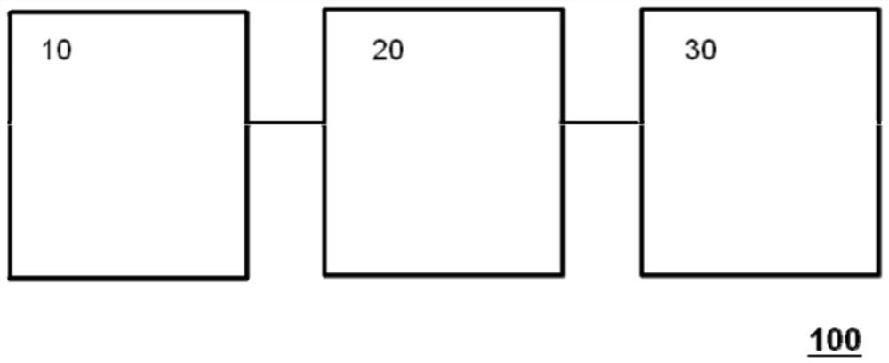
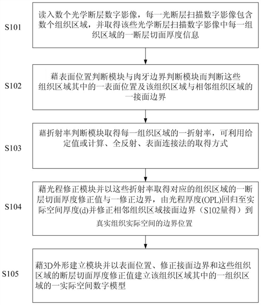
Provided are an optical tomography digital-impression imaging system and a method for use thereof, said system including: an optical coherence tomography scanner, used for obtaining tomographic slice thicknesses of a plurality of tissue areas of each optical tomography digital image; an information analysis and processing component electrically connected to the optical coherence tomography scanner. The information analysis and processing component has a refractive index compensation and optical path correction functions; by way of analysis and processing, tomographic slice thickness correction values corresponding to the tissue areas are obtained, thus establishing a digital model of the tissue areas.

Description

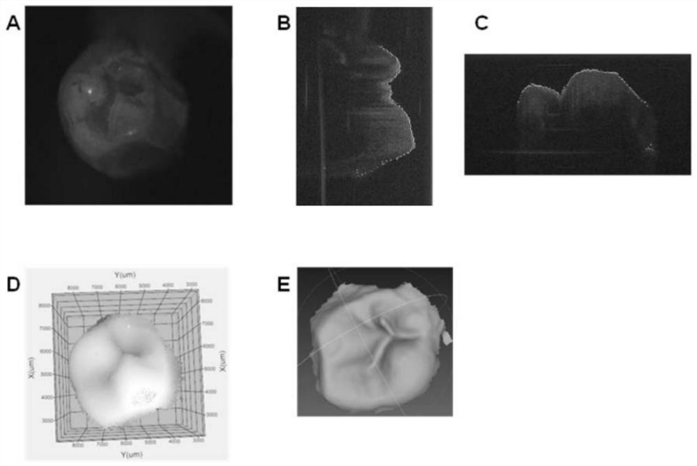
technical field [0001] The invention relates to an optical tomography digital impression image taking instrument and a using method thereof. It uses optical coherence tomography (OCT) technology to image the tooth structure, obtains the information of the tooth surface and the boundary structure of the teeth and gums under the gums, and performs tissue refractive index compensation to obtain the position of the teeth and gums in the actual space , and then establish a complete digital three-dimensional model of teeth through digital gingival retraction, and has a 3D file output function. Background technique [0002] The global fixed denture market continues to grow. To make a fixed denture, the patient's teeth must first be ground, followed by gingiva retraction to expose the ground teeth hidden in the gums, and then an impression is made. In the process, accuracy, dimensional stability, operating variables, etc. will directly affect the precision of the finished denture. ...

Claims


Application Information

Patent Timeline
Patent Type & Authority Patents(China)
IPC IPC(8): A61B5/00A61C13/00G01N21/00
CPCA61B5/0066A61B5/0073A61B5/0088A61B2576/02A61C9/0053G16H30/40
Inventor 李士元吕志伟吕东原赖昱臻
Owner FARMAR LICENSING
Learn More