
Ultrasound contrast image segmentation method based on statistical partial differential model

A technology of contrast-enhanced ultrasound and image segmentation, which is applied in image analysis, image enhancement, image data processing, etc., and can solve problems such as uneven distribution of gray levels, existence of noise, and difficulties for sonographers

Active Publication Date: 2018-05-29
THE FIRST AFFILIATED HOSPITAL OF ARMY MEDICAL UNIV +1
View PDF3 Cites 15 Cited by
  • Summary
  • Abstract
  • Description
  • Claims
  • Application Information

AI Technical Summary

Problems solved by technology

However, the result obtained by contrast-enhanced ultrasound is a video or Dicom sequence, and there are problems such as noise, uneven gray distribution, and blurred outline of the target object, which makes it difficult for sonographers to perform subjective analysis

Method used


Image

  • Ultrasound contrast image segmentation method based on statistical partial differential model
  • Ultrasound contrast image segmentation method based on statistical partial differential model
  • Ultrasound contrast image segmentation method based on statistical partial differential model

Examples


Embodiment Construction

[0060] Embodiments of the present invention are described in detail below, examples of which are shown in the drawings, wherein the same or similar reference numerals designate the same or similar elements or elements having the same or similar functions throughout. The embodiments described below by referring to the figures are exemplary only for explaining the present invention and should not be construed as limiting the present invention.

[0061] In describing the present invention, it should be understood that the terms "longitudinal", "transverse", "upper", "lower", "front", "rear", "left", "right", "vertical", The orientation or positional relationship indicated by "horizontal", "top", "bottom", "inner", "outer", etc. are based on the orientation or positional relationship shown in the drawings, and are only for the convenience of describing the present invention and simplifying the description, rather than Nothing indicating or implying that a referenced device or elem...


PUM


Abstract

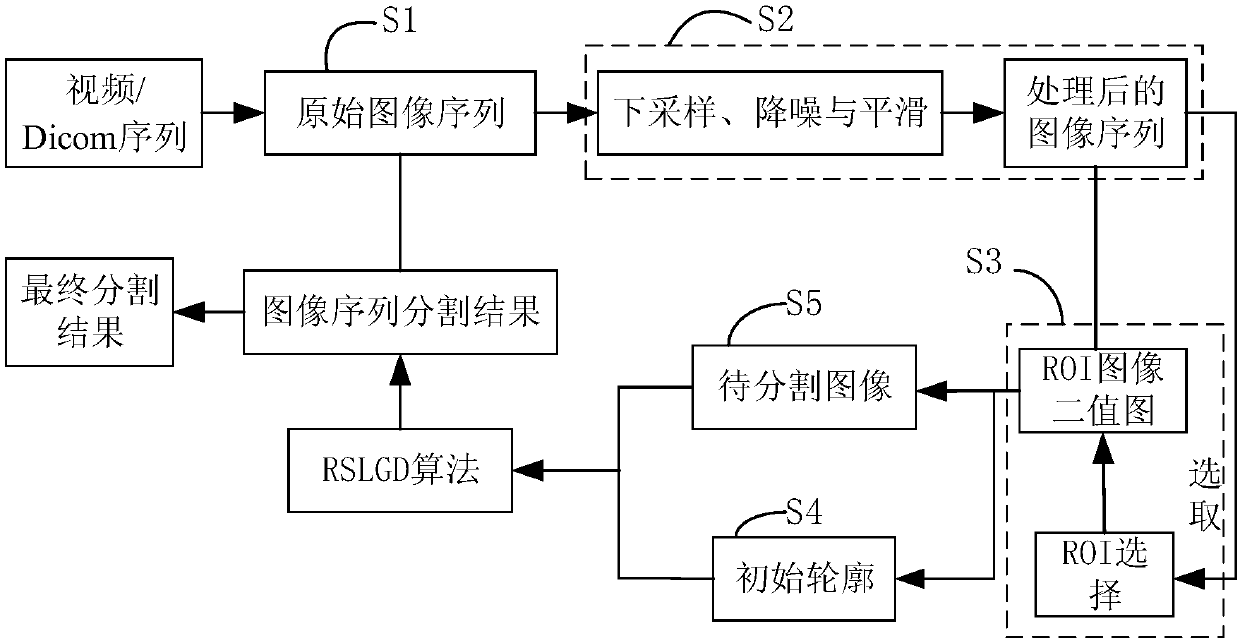
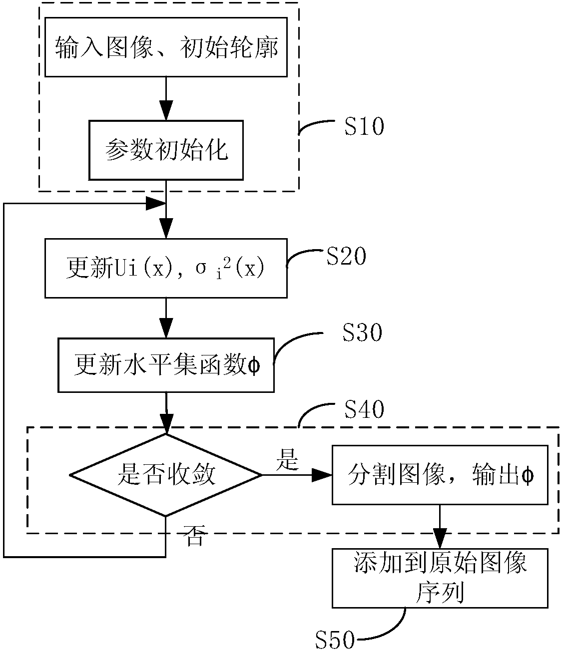
The invention discloses an ultrasound contrast image segmentation method based on a statistical partial differential model. The method comprises a preprocessing step and an online segmentation step. At the preprocessing step, a to-be-segmented image and an initial contour are obtained; and at the online segmentation step, an energy function is obtained by using the to-be-segmented image and the initial contour as inputs, an edge stop function is introduced into the energy function to constrain the length item and an LoG image energy function is added, numerical solution is carried out on the energy function, and target edge capturing and segmentation are carried out on a single or a plurality of different ROI images quickly. The ROI in an ultrasound contrast image is segmented; a doctor isassisted in obtaining time strength curves of masses in a lesion region and same level reference regions like parenchymal organs including the liver and the kidney and the like and superficial organsincluding the breast and the thyroid gland and the like by observing the segmentation result of the whole sequence, so that a quantitative index is obtained by the analysis.

Description

technical field [0001] The invention relates to a method for segmenting contrast-enhanced ultrasound images, in particular to a method for segmenting contrast-enhanced ultrasound images based on a statistical partial differential model. Background technique [0002] Image segmentation technology occupies a very important position in the qualitative and quantitative analysis of medical contrast-enhanced ultrasound images. In the application of clinical contrast-enhanced ultrasonography, sonographers often judge the blood of the lesion by subjectively analyzing the perfusion intensity, uniformity, and perfusion mode (diffuse enhancement / central enhancement) of different dynamic images of Region of Interest (ROI). For the situation. However, the result obtained by CEUS is a video or Dicom sequence, and there are problems such as noise, uneven gray level distribution, and blurred outline of the target object, which makes it difficult for sonographers to perform subjective analy...

Claims


Application Information

Patent Timeline
Patent Type & Authority Applications(China)
IPC IPC(8): G06T7/11G06T7/12G06T7/13G06T7/136G06T7/149
CPCG06T7/11G06T7/12G06T7/13G06T7/136G06T7/149G06T2207/10132
Inventor 郭燕丽房斌张伟王翊谭立文方可敬朱连华刘灯
Owner THE FIRST AFFILIATED HOSPITAL OF ARMY MEDICAL UNIV
Learn More