Vascular Image Segmentation Method Based on Centerline Extraction, Magnetic Resonance Imaging System

A blood vessel image and centerline technology, applied in the field of nuclear magnetic resonance imaging systems, can solve the problems of time-consuming and labor-intensive manual designation of labels, affecting computing efficiency, and low segmentation accuracy, achieving good learning ability, improving computing efficiency, and improving the effect of segmentation efficiency.

Active Publication Date: 2020-12-29
NORTHWEST UNIV
View PDF2 Cites 0 Cited by
  • Summary
  • Abstract
  • Description
  • Claims
  • Application Information

AI Technical Summary

Problems solved by technology

[0003] To sum up, the problems existing in the existing technology are: the current blood vessel segmentation method is sensitive to noise, and the segmentation accuracy is low; manually specifying the label is time-consuming and laborious, which affects the calculation efficiency

Method used


Image

  • Vascular Image Segmentation Method Based on Centerline Extraction, Magnetic Resonance Imaging System
  • Vascular Image Segmentation Method Based on Centerline Extraction, Magnetic Resonance Imaging System
  • Vascular Image Segmentation Method Based on Centerline Extraction, Magnetic Resonance Imaging System

Examples


Embodiment Construction

[0046] In order to make the object, technical solution and advantages of the present invention more clear, the present invention will be further described in detail below in conjunction with the examples. It should be understood that the specific embodiments described here are only used to explain the present invention, not to limit the present invention.

[0047] The invention realizes the segmentation of cerebral blood vessels, and has the characteristics of accuracy, speed and no human intervention. Its true positive rate and true negative rate can reach 0.85, and the segmentation accuracy has been improved to a certain extent compared with the existing technology.

[0048] The application principle of the present invention will be described in detail below in conjunction with the accompanying drawings.

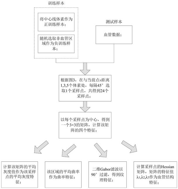
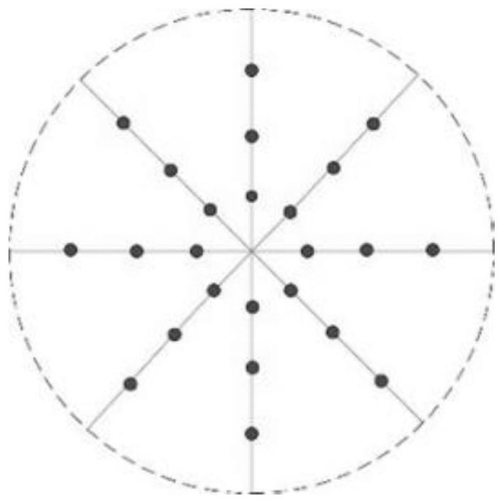
[0049] Such as figure 1 As shown, the blood vessel image segmentation method based on centerline extraction provided by the embodiment of the present invention includes ...


PUM


Abstract

The invention belongs to the technical field of medical image processing, and discloses a blood vessel image segmentation method based on centerline extraction, a nuclear magnetic resonance imaging system, and preprocessing of brain blood vessel data by vesselness filtering based on Hessian matrix; topology refinement method for blood vessel centerline Extraction; take the centerline point as a positive sample and the non-vascular point as a negative sample to extract the features of the training sample and the test sample; use the features of the training sample and the corresponding label to train the SVM model, and use the feature of the test sample as the input of the trained SVM model , the output label is the segmentation result of blood vessels. The invention reduces the workload and improves the calculation efficiency; it does not need to manually calibrate the target and the background, completes automatic blood vessel segmentation, and greatly improves the segmentation efficiency. The invention realizes the segmentation of cerebral blood vessels, which is accurate, fast and does not require human intervention; the true positive rate and true negative rate can reach 0.85.

Description

technical field [0001] The invention belongs to the technical field of medical image processing, and in particular relates to a blood vessel image segmentation method based on central line extraction and a nuclear magnetic resonance imaging system. Background technique [0002] Vessel segmentation is one of the most important medical image processing techniques, which is crucial to the diagnosis and treatment of cardiovascular and cerebrovascular diseases and other related diseases. Accurate segmentation is the primary issue of image analysis and recognition, and it is also a factor that restricts the development and application of other related technologies, such as blood vessel matching, 3D reconstruction, and motion estimation. Due to the influence of imaging noise, complex vascular structure, and other factors, medical images usually have low contrast and blurred boundaries between different tissues, while fine structures like blood vessels are susceptible to noise and n...

Claims


Application Information

Patent Timeline
Patent Type & Authority Patents(China)
IPC IPC(8): G06T7/10G06T7/68G06K9/62
Inventor 侯榆青孙飞飞赵凤军贺小伟陈一兵高培王宾易黄建曹欣
Owner NORTHWEST UNIV
Why Eureka
Try Eureka