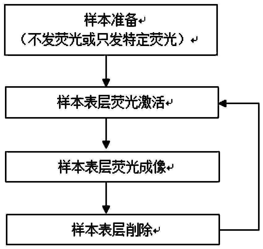
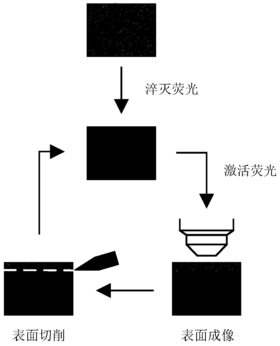
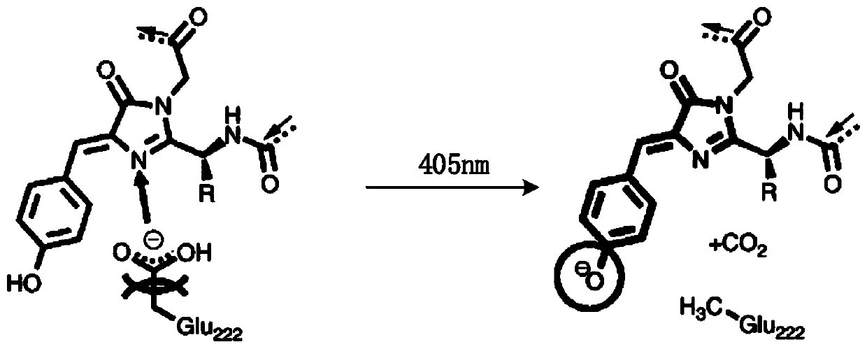
A method of tomographic imaging
A tomography and imaging technology, which is applied in the field of biological fluorescence microscopy imaging, can solve the problems of requirements, limitations, and high sample transparency of light sheet lighting technology, and achieve shortened imaging time, fast imaging speed, and good tomographic effect Effect
- Summary
- Abstract
- Description
- Claims
- Application Information
AI Technical Summary
Problems solved by technology
Method used
Image
Examples
Embodiment 1
[0088] Chemical tomography of the whole brain of Thy1-EGFP-M mice, including the following steps:
[0089] (1) The sample is fixed.
[0090] The whole brain of Thy1-EGFP-M mice was fixed by means of chemical fixation to obtain fixed mouse brain biological tissue. Specific steps are as follows:
[0091] After the heart was perfused at 4°C, the dissected whole brain of the mouse was soaked in 4% PFA solution for about 12 hours. Use 40ml of PBS solution, rinse for four hours each time.
[0092] (2) Sample dehydration.
[0093] The fixed mouse brain tissue was replaced with ethanol, so that the biological tissue was dehydrated, and the dehydrated EGFP-labeled mouse whole brain was obtained. The specific steps are:
[0094] At 4°C, dehydrate the fixed mouse whole brain sequentially through 20 ml of gradient ethanol double-distilled aqueous solution for 2 hours. The concentration gradient of ethanol double distilled water solution is 50%, 75%, 95%, 100%, 100% ethanol according...
Embodiment 2
[0105] A method for chemical tomography of the mouse whole brain overexpressing the pH-sensitive fluorescent protein pHuji, comprising the following steps:
[0106] (1) The sample is fixed.
[0107] The whole brain of mice overexpressing pHuji was fixed by chemical fixation means to obtain fixed pHuji-labeled biological tissues. The specific steps are as follows: at 4 degrees Celsius, after perfusing the heart, the dissected whole brain of the mouse was soaked in a 4% PFA solution for about 12 hours, the amount of PFA solution was 20ml per mouse, and then rinsed three times with PBS solution. Each animal uses 40ml of PBS solution each time, and rinses for four hours each time.
[0108] (2) Sample dehydration.
[0109]The fixed pHuji-labeled mouse whole brain was replaced with ethanol, so that the biological tissue was dehydrated, and the dehydrated pHuji-labeled mouse whole brain was obtained. The specific steps are as follows: at 4°C, soak the fixed pHuji-labeled mouse who...
Embodiment 3
[0120] A chemical tomography method for the whole brain of mice overexpressing EYFP, comprising the following steps:
[0121] (1) The sample is fixed.
[0122] The whole brain of the overexpressing EYFP mouse was fixed by chemical fixation means to obtain the fixed mouse brain biological tissue. The specific steps are as follows: at 4 degrees Celsius, after perfusing the heart, the dissected whole brain of the mouse was soaked in a 4% PFA solution for about 12 hours, the amount of PFA solution was 20ml per mouse, and then rinsed three times with PBS solution. Each animal uses 40ml of PBS solution each time, and rinses for four hours each time.
[0123] (2) Sample dehydration.
[0124] The fixed mouse brain tissue was replaced with ethanol, so that the biological tissue was dehydrated, and the dehydrated EGFP-labeled mouse whole brain was obtained. The specific steps are as follows: at 4 degrees Celsius, the fixed mouse whole brain is sequentially passed through 20 ml of gra...
PUM
| Property | Measurement | Unit |
|---|---|---|
| thickness | aaaaa | aaaaa |
Abstract
Description
Claims
Application Information
 - R&D
- Intellectual Property
- Life Sciences
- Materials
- Tech Scout
- Unparalleled Data Quality
- Higher Quality Content
- 60% Fewer Hallucinations
Browse by: Latest US Patents, China's latest patents, Technical Efficacy Thesaurus, Application Domain, Technology Topic, Popular Technical Reports.
© 2025 PatSnap. All rights reserved.Legal|Privacy policy|Modern Slavery Act Transparency Statement|Sitemap|About US| Contact US: help@patsnap.com


