
Thoracic cavity simulator

A technology of simulator and thoracic cavity, applied in the field of thoracic cavity simulator, can solve the problems that the thoracic cavity simulator has not yet appeared

Active Publication Date: 2017-02-22
FASOTEC +1
View PDF7 Cites 5 Cited by
  • Summary
  • Abstract
  • Description
  • Claims
  • Application Information

AI Technical Summary

Problems solved by technology

[0006] At present, there is no thoracic cavity simulator that can faithfully reproduce the shape or texture of the human body and simulate the human surgical environment with many limitations for training or learning of thoracoscopic surgery.

Method used


Image

  • Thoracic cavity simulator
  • Thoracic cavity simulator
  • Thoracic cavity simulator

Examples


Embodiment 1

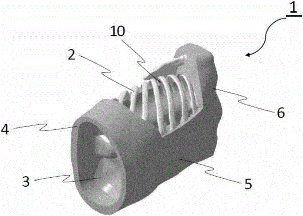
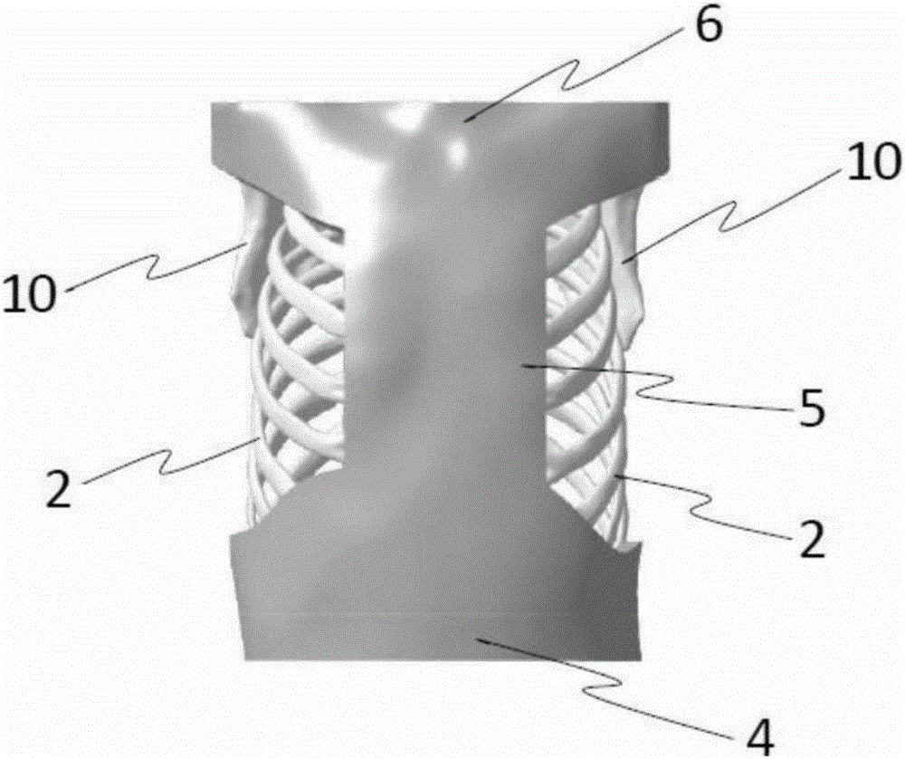
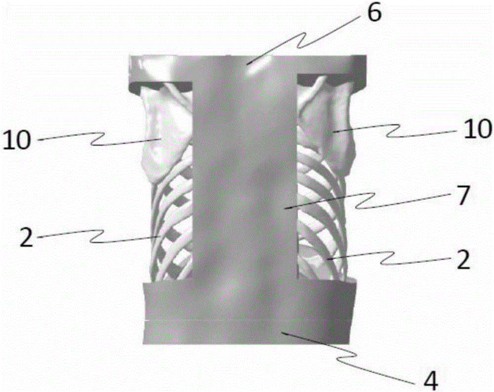
[0052] figure 1 Shown is an external view of the thorax simulator 1 . Figure 2-7 They are the front view, rear view, right side view, left side view, plan view and bottom view of the chest cavity simulator 1 respectively.

[0053] Chest simulator 1 is composed of a human skeleton model simulating ribs 2, sternum, spine and scapula 10, and a housing (4, 5, 6) for accommodating the human skeleton model. The rib portion of the housing is provided with an opening. Bottom cover 4 with convex part simulating diaphragm 3 can be freely attached to and detached from the case, can be opened and closed on the diaphragm part, and can store organ models such as lungs or heart inside the human skeleton model of ribs . The openings on the rib portion of the casing expose the second to eighth ribs, and the openings are provided on the left side and the right side, respectively.

[0054] The human skeleton model of sternum is hidden in the inner side of housing front part 5. In addition,...


PUM


Abstract

Provided is a thoracic cavity simulator that, for the purpose of training or education in thoracic cavity microscopic surgery, faithfully reproduces the shape and feel of a human body and that can simulate a surgical environment for a human body that has multiple constraints. A device that comprises a model human skeleton that simulates at least ribs, and comprises a casing that houses the model human skeleton, the device being configured such that an opening is provided to a rib section of the casing, such that a diaphragm section can be opened and closed, and such that model organs can be housed inside the ribs of the model human skeleton. The shapes / structures and hardness / feel, etc. of the surfaces and interiors of the model organs inside the ribs of the model human skeleton are made to approximate a human body. The casing may have a hardness / feel, etc. that approximates a human body or may be a hard resin. An opening is provided to the rib section of the casing, and forceps used for thoracic cavity microscopic surgery are inserted inside the ribs through gaps between rib bones. The diaphragm section is configured so as to be removable and / or openable and closable, and the model organs housed inside the ribs of the model human skeleton are replaced.

Description

technical field [0001] The invention relates to a thoracic cavity simulator used for training or learning thoracoscopic operation skills. Background technique [0002] In the medical field, the need for three-dimensional visualization of the affected part or specific parts of the body is increasing, such as signing informed consent, deciding on treatment plans, medical education, and medical research. Especially when using 3D modeling molds to achieve 3D visualization, not only vision, but also actual contact observation of three-dimensional shapes with hands can obtain more information that cannot be conveyed by computer images. [0003] In recent years, three-dimensional printers have been disclosed that combine hard and soft resins by injecting multiple resins at the same time, and create three-dimensional modeling molds using resins with different mechanical properties. Using a 3D printer, the surface associated with the shape structure, its internal structure can also ...

Claims


Application Information

Patent Timeline
IPC IPC(8): G09B23/34G09B9/00
CPCG09B23/34G09B9/00G09B23/285
Inventor 渡边欣一安乐武志森川利昭
Owner FASOTEC
Learn More