Method and system for hemorrhage area segmentation in brain CT images based on semi-supervised learning

A semi-supervised learning and CT image technology, applied in image analysis, image enhancement, graphics and image conversion, etc., can solve the problems of ignoring inter-frame information and poor effect, and achieve the effect of simple processing method and easy extraction

Active Publication Date: 2019-02-01
ZHEJIANG UNIV
View PDF2 Cites 1 Cited by
  • Summary
  • Abstract
  • Description
  • Claims
  • Application Information

AI Technical Summary

Problems solved by technology

First, most of these methods use very simple segmentation algorithms, such as clustering and thresholding, etc., and while these methods may perform well in natural image processing, they do not perform well in complex situations, such as bleeding regions overlapping brain tissue. These methods do not work well when the edges of the bleed or bleed are not sufficiently discriminative
Second, most of the existing algorithms are only suitable for processing two-dimensional images
However, CT imaging is a three-dimensional process, so a series of parallel scanning image frames will be generated, while the 2D segmentation algorithm will ignore some important inter-frame information

Method used


Image

  • Method and system for hemorrhage area segmentation in brain CT images based on semi-supervised learning
  • Method and system for hemorrhage area segmentation in brain CT images based on semi-supervised learning
  • Method and system for hemorrhage area segmentation in brain CT images based on semi-supervised learning

Examples


Embodiment Construction

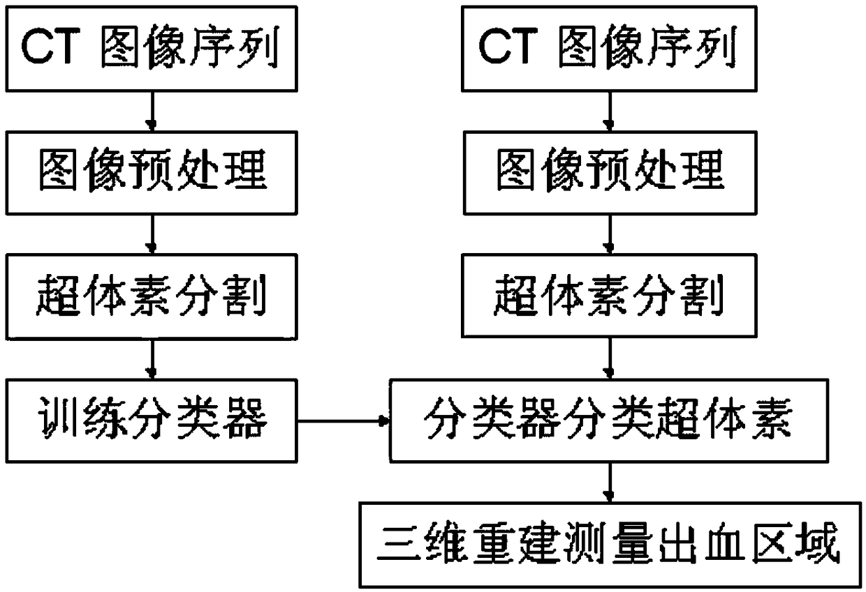
[0061] The invention is applicable to the hemorrhage area segmentation in the medical cranial CT image, and is a method for segmenting the hemorrhage area of ​​the brain CT image based on semi-supervised learning and three-dimensional supervoxel.

[0062] The flow chart of the present invention is as figure 1 , mainly including the Tri-training model training stage and the bleeding area segmentation stage based on the Tri-training model.

[0063] The Tri-training model training phase includes the following steps:

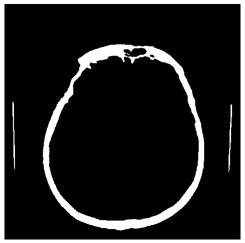
[0064] (1.1) Converting the CT image format: Obtain the CT image sequence containing the hemorrhage area from the computer tomography equipment or database, intercept the effective interval of the pixel value, and convert it into a commonly used computer image processing format. figure 2 That is, the image obtained after the format conversion of the CT image.

[0065] (1.2) Mark training samples: Divide the CT image sequence into two parts, one part of the sequen...


PUM


Abstract

The invention discloses a method for segmenting bleeding areas in brain CT images based on semi-supervised learning. The method includes a semi-supervised model training stage and a semi-supervised model-based bleeding area segmentation stage; the semi-supervised model training stage is used to train semi-supervised model; the bleeding area segmentation stage based on the semi-supervised model includes format conversion of the two-dimensional CT image sequence that needs to be segmented into the intracranial hemorrhage area, reconstructing the two-dimensional CT image into a three-dimensional space, and then using the supervoxel algorithm to divide the three-dimensional image into For supervoxels of similar size, features are extracted from each supervoxel as a sample, and finally the supervoxels are divided into foreground and background parts through a trained semi-supervised model based on the features. The present invention effectively improves the accuracy of bleeding area detection by introducing a semi-supervised learning algorithm and using supervoxels instead of pixels for calculation.

Description

technical field [0001] The present invention relates to the fields of machine learning and image processing, in particular to a method and system for segmenting hemorrhage areas in brain CT images based on semi-supervised learning. Background technique [0002] Intracranial hemorrhage (ICH) is one of the most serious acute cerebrovascular diseases, and it is also an important cause of acute neurological disorders such as hemiplegia. Therefore, for clinical treatment, early diagnosis of intracranial hemorrhage is of great significance. Compared with clinical manifestations, computed tomography (CT) scan and magnetic resonance imaging (MRI) scan, which can be detected without trauma, can more directly and accurately reflect the severity and evolution trend of intracranial hemorrhage. At the same time, because the cost of CT detection is much less than that of MRI detection, most patients will choose CT detection. Fresh hematomas usually appear as hyperluminous areas with ind...

Claims


Application Information

Patent Timeline
IPC IPC(8): G06T7/11G06T17/00G06T3/00
CPCG06T2207/10081G06T2207/20084G06T2207/20081G06T3/06
Inventor 胡浩基孙明杰
Owner ZHEJIANG UNIV
Why Eureka
Try Eureka