Femur segmenting method in tomographic image

A technology of tomography and femur, applied in the field of image processing, to achieve the effect of solving inaccurate segmentation

Active Publication Date: 2016-10-12
FUJIAN UNIV OF TECH
View PDF8 Cites 5 Cited by
  • Summary
  • Abstract
  • Description
  • Claims
  • Application Information

AI Technical Summary

Problems solved by technology

This method will consume a lot of labor costs, and has certain requirements for the operator's experience

Method used


Image

  • Femur segmenting method in tomographic image
  • Femur segmenting method in tomographic image
  • Femur segmenting method in tomographic image

Examples


Embodiment 1

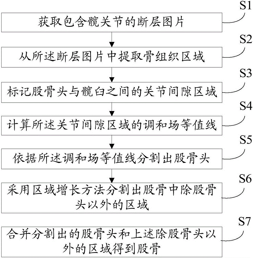
[0065] Please refer to Figure 2 to Figure 9 , Embodiment 1 of the present invention is:

[0066] A method for segmenting a femur in a tomographic image, comprising:

[0067] S1, obtain a plurality of cross-sectional pictures comprising the hip joint; the hip joint comprises the femoral head and the acetabulum, and the femoral head is connected with the femoral neck, the greater trochanter, the lesser trochanter, etc. in the femur; therefore, the cross-sectional pictures comprising the hip joint are at least Including the femoral head and the area of ​​the femur other than the femoral head;

[0068] Specifically, load tomographic image data from DICOM and other format files, and perform visualization processing through a computer graphics visualization platform (such as OpenGL), obtain multi-layer tomographic images, and display them on a visual interactive interface;

[0069] S2. Extract the bone tissue area from the tomographic image;

[0070] Specifically, use the histog...


PUM


Abstract

The invention provides a femur segmenting method in a tomographic image. The method comprises the steps that a tomographic picture with a hip joint is acquired; a bone tissue region is extracted from the tomographic picture; a joint gap region between femoral head and acetabulum is marked; the harmonic field contour of the joint gap region is calculated; and femoral head is segmented according to the harmonic field contour. The joint gap region is marked to acquire the approximate range of the joint gap region. The harmonic field contour of the joint gap region is calculated to acquire the exact joint gap region boundary and the femoral head boundary. Femoral head is efficiently and accurately segmented. The degree of automation is high. The femur segmenting method has an important significance for the assessment of joint wear, hip arthritis auxiliary diagnosis, hip replacement surgery simulation and other follow-up applications.

Description

technical field [0001] The invention relates to the field of image processing, in particular to a method for segmenting a femur in a tomographic image. Background technique [0002] The hip joint is one of the important joints of the human body. It is mainly composed of a spherical femoral head, a socket-shaped acetabulum, and ligaments and cartilage between the two; The greater and lesser trochanters connected by bones, the femoral neck and other tissues. On the other hand, computerized tomography technology, commonly known as CT technology, can reveal tissue information invisible to the naked eye through the skin tissue. Therefore, as a computer-aided technology, CT technology is widely used in clinical medicine; for example Evaluation of joint tissue wear, auxiliary diagnosis of hip arthritis, simulation of hip replacement surgery, etc. [0003] A tomographic picture can be understood as a series of equally spaced tissue cross-sectional pictures produced by scanning a s...

Claims


Application Information

Patent Timeline
Patent Type & Authority Applications(China)
IPC IPC(8): G06T7/00
CPCG06T2207/10081G06T2207/30008
Inventor 刘石坚邹峥罗三定廖胜辉潘正祥邹复民廖律超聂明星杨海燕
Owner FUJIAN UNIV OF TECH
Why Eureka
Try Eureka