
Ventricular premature beat abnormal activation site positioning method based on ECGI (electrocardiographic imaging)

A premature ventricular contraction and point positioning technology, applied in 3D image processing, medical science, image data processing, etc., can solve problems such as low efficiency

Active Publication Date: 2016-07-27
ZHEJIANG UNIV
View PDF12 Cites 14 Cited by
  • Summary
  • Abstract
  • Description
  • Claims
  • Application Information

AI Technical Summary

Problems solved by technology

However, invasive methods are not efficient and have certain risks

Method used


Image

  • Ventricular premature beat abnormal activation site positioning method based on ECGI (electrocardiographic imaging)
  • Ventricular premature beat abnormal activation site positioning method based on ECGI (electrocardiographic imaging)
  • Ventricular premature beat abnormal activation site positioning method based on ECGI (electrocardiographic imaging)

Examples


Embodiment Construction

[0026] In order to describe the present invention more clearly, the technical solution of the present invention will be described in detail below in conjunction with the accompanying drawings and specific embodiments.

[0027] The present invention is based on the method for locating abnormal premature ventricular beats excited points based on ECGI, and the specific implementation steps are as follows:

[0028] S1. Acquisition of 64-lead body surface potential data and thoracic computed tomography imaging data of a patient with premature ventricular contraction wearing a body surface potential recording vest.

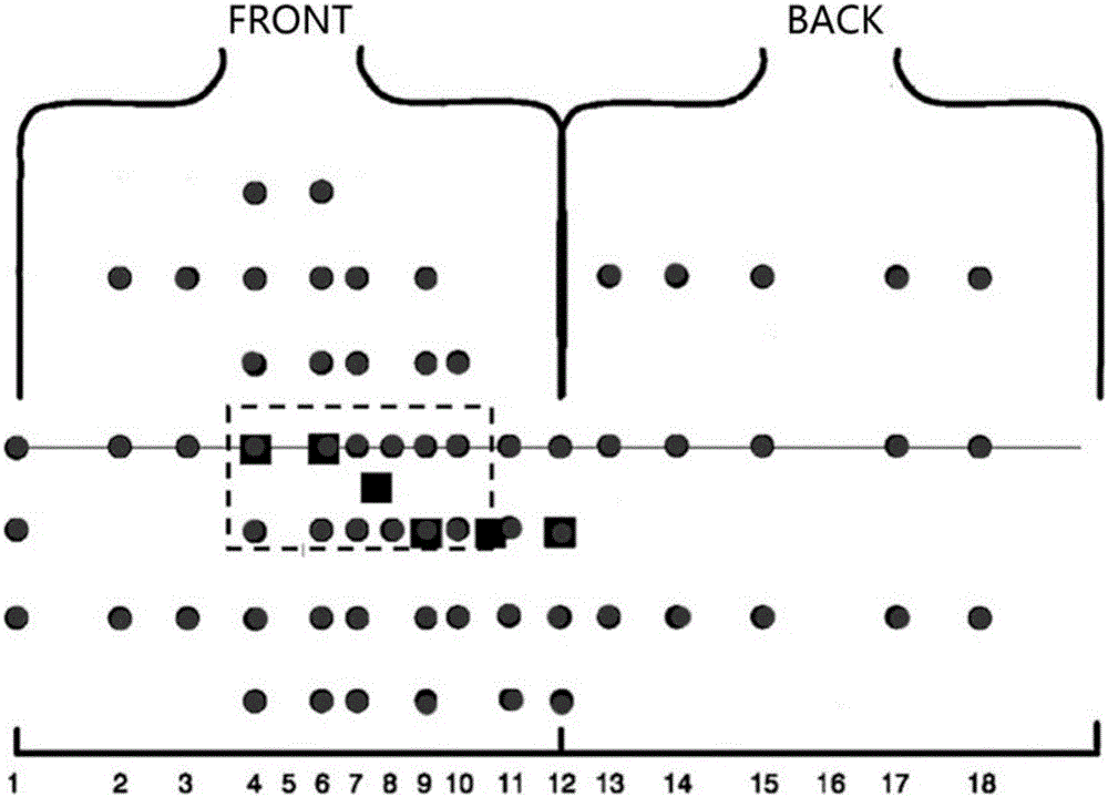
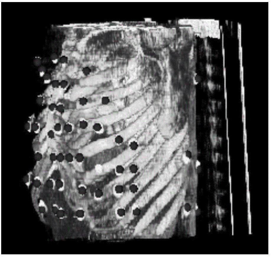
[0029] The patient first wears a vest with 64 electrodes to record the body surface potential data. The distribution of the 64 leads on the body surface is as follows: figure 1 shown; the patient then underwent computed tomography while wearing the vest after removing or shortening the leads from the vest, recording the geometry of the torso and heart.

[0030] S2. Bui...


PUM


Abstract

The invention discloses a ventricular premature beat abnormal activation site positioning method based on ECGI (electrocardiographic imaging). According to the method, by establishing a combined collection framework of 64 lead body surface potential data and computed tomography imaging, a personalized heart-trunk model is obtained; by Tikhonov regularization, the ECG (Electrocardiograph) inverse problem is solved; an epicardial potential is reconstructed so as to accurately position a ventricular premature beat abnormal activation site. The ventricular premature beat abnormal activation site positioning method has important actual application value.

Description

technical field [0001] The invention belongs to the technical field of cardiac electrophysiological analysis, and in particular relates to an ECGI-based method for locating abnormal premature ventricular beat activation points. Background technique [0002] Electrocardiography (Electrocardiography) has become a very important method for non-invasively revealing cardiac electrophysiological activities by recording the potential changes caused by the depolarization and repolarization process during the cardiac beating cycle on the body surface. If the heart is regarded as the source of the electric field, then the corresponding electric field is distributed in the space from the heart to the body surface, and the potentials at various positions can be recorded on the body surface. The so-called electrophysiological inversion of the heart is to infer from the outside to the inside the abnormality of the electrical activity in the heart caused by myocardial ischemia through the ...

Claims


Application Information

Patent Timeline
IPC IPC(8): A61B5/0402A61B5/0432G06T15/10G06T5/00
CPCG06T15/10A61B5/7203A61B5/725G06T2215/06A61B5/316A61B5/333A61B5/318G06T5/80G06T5/70
Inventor 刘华锋马骁勇
Owner ZHEJIANG UNIV
Learn More