
Imaging system of endoscope

An imaging system and endoscope technology, applied in endoscopy, medical science, sensors, etc., can solve the problems of shallow light projection depth, inability to judge blood vessel obstruction, etc., and achieve the effect of improving the recognition ability

Active Publication Date: 2016-07-13
SONOSCAPE MEDICAL CORP
View PDF7 Cites 20 Cited by
  • Summary
  • Abstract
  • Description
  • Claims
  • Application Information

AI Technical Summary

Problems solved by technology

[0005] The purpose of the present invention is to provide an endoscopic imaging system, which aims to solve the problem that the light projection depth is shallow in the existing narrow bandwidth imaging technology, and only the absorption characteristics of superficial tissue can be obtained, and it is impossible to judge whether the blood vessel is blocked

Method used


Image

  • Imaging system of endoscope
  • Imaging system of endoscope
  • Imaging system of endoscope

Examples


Embodiment Construction

[0023] In order to enable those skilled in the art to better understand the solution of the present invention, the present invention will be further described in detail below in conjunction with the accompanying drawings and specific embodiments. Apparently, the described embodiments are only some of the embodiments of the present invention, but not all of them. Based on the embodiments of the present invention, all other embodiments obtained by persons of ordinary skill in the art without making creative efforts belong to the protection scope of the present invention.

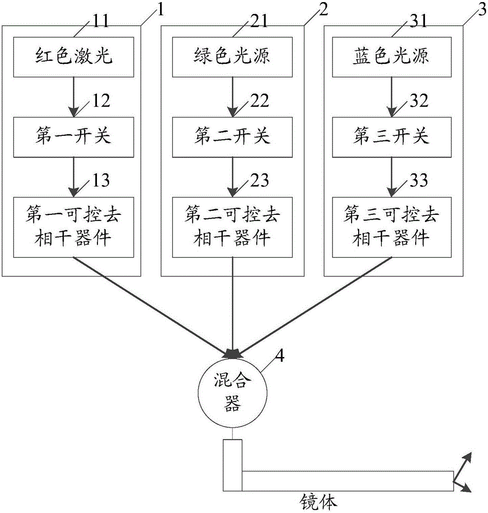
[0024] A structural block diagram of a specific embodiment of the endoscopic imaging system provided by the present invention is as follows: figure 1 As shown, the device includes:

[0025] Red laser 11, green light source 21, blue light source 31, first switch 12, second switch 22, third switch 32, first controllable decoherence device 13, second controllable decoherence device 23, third controllable Decohe...


PUM


Abstract

The invention discloses an imaging system of an endoscope.According to the system, one end of a first switch is connected with red laser and the other end of the first switch is connected with a first controllable decorrelation device to serve as a first channel; one end of a second switch is connected with a green light source and the other end of the second switch is connected with a first controllable decorrelation device to serve as a second channel; one end of a third switch is connected with a blue light source and the other end of the third switch is connected with a third controllable decorrelation device to serve as a third channel; the first channel, the second channel and the third channel are connected with a mixer, and mixed signal light is output; the first controllable decorrelation device in the first channel is not enabled, output signal light is used for laser speckle imaging, a laser speckle angiography image is generated, output signal light of the second channel and the third channel is used for narrow-band light imaging, and a narrow-band light image is generated.The imaging system of the endoscope breaks through narrow-band light imaging depth, detects the angiemphraxis condition, and improves the capacity of recognizing blood vessel depth information.

Description

technical field [0001] The invention relates to the technical field of endoscope clinical function imaging, in particular to an endoscope imaging system. Background technique [0002] With the development of science and technology, a series of new endoscopic techniques are applied clinically. The NBI narrow-band imaging developed by Olympus Medical Systems Corporation of Japan has created an unprecedented advantage for the early detection of patients with nasopharyngeal tumors. [0003] In the human body, the main pigment of mucosal tissue is hemoglobin. Hemoglobin has a peak absorption capacity for blue light and relatively weak absorption for green light. Compared with conventional endoscopes, NBI technology uses narrow-band filters to filter out red light, leaving green light and blue light with center wavelengths of 540nm and 415nm respectively. Since the narrow-wavelength light does not cover all the visible light spectrum, the depth of reaching the mucosa is differe...

Claims


Application Information

Patent Timeline
IPC IPC(8): A61B1/06A61B5/026
CPCA61B1/0638A61B5/0261
Inventor 陈云亮冯能云邱建军
Owner SONOSCAPE MEDICAL CORP
Learn More