
Method for isolating tumor cell-derived exosomes from malignant pleural effusion

A technology for malignant pleural effusion and tumor cells, applied in tumor/cancer cells, cell dissociation methods, animal cells, etc., can solve the problems of restricting researchers' research, complicated and time-consuming steps, complex equipment requirements, etc., to overcome tumor specificity. Low performance, simple and effective cost, accurate and reliable method

Active Publication Date: 2018-09-28
ZHEJIANG CANCER HOSPITAL
View PDF4 Cites 0 Cited by
  • Summary
  • Abstract
  • Description
  • Claims
  • Application Information

AI Technical Summary

Problems solved by technology

[0005] Exosomes can also be released by tumor cells in pleural effusions, but why they are released by tumors into the pleura and what role they play is unclear
Existing methods for extracting exosomes from pleural effusion are complicated and require high equipment. For example, the literature reports a method for extracting exosomes from pleural effusion by sucrose gradient ultracentrifugation. , but also need to configure ultra-high-speed centrifuges, which greatly limit the researcher's research
In addition, the origin of exosomes in pleural effusion is complex, and it is impossible to assess the exosomes secreted by tumor cells
Based on the above limitations, there are no published reports on methods for isolating tumor cell-derived exosomes from pleural effusions.

Method used


Image

  • Method for isolating tumor cell-derived exosomes from malignant pleural effusion
  • Method for isolating tumor cell-derived exosomes from malignant pleural effusion
  • Method for isolating tumor cell-derived exosomes from malignant pleural effusion

Examples


Embodiment 1

[0039] Example 1: Extraction of exosome derived from tumor cells in pleural effusion of lung cancer patients

[0040] ①With the patient’s informed and ethical consent, collect 2ml of preoperative pleural effusion specimens from tumor patients (pathological biopsy has confirmed lung cancer) in the first 10ml centrifuge tube, and centrifuge at 4°C and 300g for 10 minutes , separated to remove the cells in the pleural effusion, and then to obtain the supernatant fluid.

[0041] ② Transfer the upper layer liquid obtained in ① into a second centrifuge tube, centrifuge at 4°C and 10,000 g for 15 minutes, separate to remove organelles and other impurities, and obtain a supernatant.

[0042] ③ Transfer the supernatant into the third centrifuge tube, add the same amount of exosome extract (Total Exosome Isolation, Invitrogen) as the volume of the supernatant, mix, shake evenly, and incubate at 4°C overnight (6-16 hours average can), and then, centrifuge the incubated mixture at 4° C. ...

Embodiment 2

[0071] Example 2: Extraction of tumor-derived exosome in pleural effusion of patients with esophageal cancer

[0072] The tumor-derived exosome in the pleural effusion of a patient with esophageal cancer was extracted by basically the same method as in Example 1. That is: ① The pleural effusion sample in the middle is a 5ml pleural effusion sample from a patient with esophageal cancer before operation, and other steps are exactly the same as in Example 1.

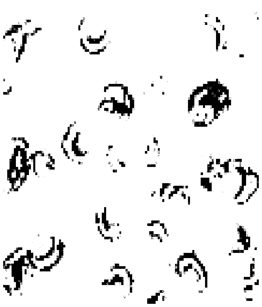
[0073] Similarly, the method in Example 1 was used to observe the product obtained in Example 2 with an electron microscope, and the morphology of exosomes consistent with the literature was also observed.

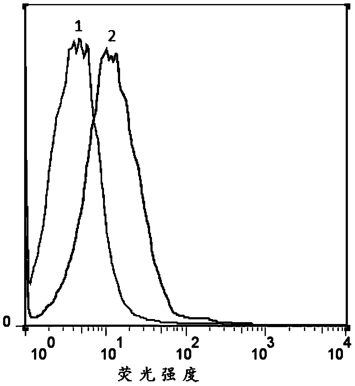
[0074] Further, immunofluorescence and flow cytometry were used to detect CD63 expression:

[0075] Resuspend the product obtained in Example 2 (i.e., tumor cell-derived exosome bound to magnetic beads) with 1 ml of 1×PBS to obtain a tumor exosome resuspension, that is, 1 ml of pleural effusion tumor-derived exosome re...

Embodiment 3

[0078] Example 3: Extraction of tumor-derived exosomes in the pleural effusion of ovarian cancer patients

[0079] The tumor-derived exosome in the pleural effusion of a patient with ovarian cancer was extracted by basically the same method as in Example 1. That is: ① The pleural effusion sample in the middle is 2.5ml of the preoperative pleural effusion sample of an ovarian patient, and the other steps are exactly the same as those in Example 1.

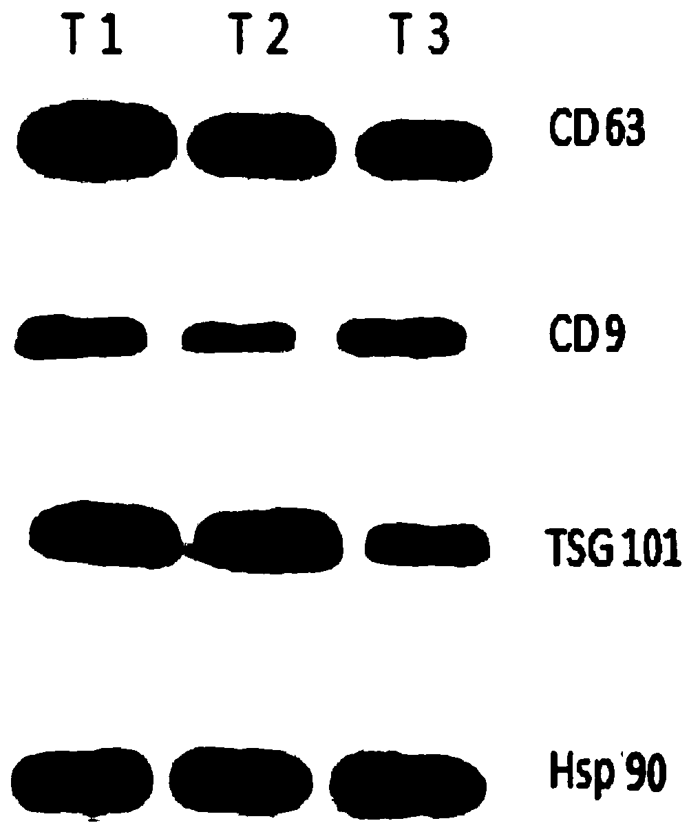
[0080] Using the product characterization method in Example 1, the product obtained in Example 3 was observed and tested. The results showed that: the product obtained in Example 3 had an exosome morphology that was consistent with that recorded in the literature, and the unique characteristics of exosomes were detected in the product. The expression of proteins CD9, CD63, Hsp90, Tsg101 and the strong expression of tumor antigen EpCAM protein prove that the product obtained in Example 3 is the exosome derived from tumor cells extrac...


PUM


Abstract

The invention discloses a method separating tumor cell-derived exosome from malignant pleural effusion, which comprises the steps in sequence: performing decellularization on a malignant pleural effusion sample, then using an extracting reagent to extract total exosome, and then using an EpCAM antibody-labeled magnetic bead to adsorb to separate and purify the tumor cell-derived exosome from the malignant pleural effusion. The method is simple and feasible and firstly realizes the extracting of the tumor cell-derived exosome in the malignant pleural effusion; the method is convenient and efficient and obtains the tumor cell-derived exosome, of which the concentration can be detected, in the malignant pleural effusion; the method is accurate and reliable, overcomes the defect of low tumor specificity of the existing total exosome detection and realizes the aim of accurately detecting the tumor cell-derived exosome biological information in the malignant pleural effusion, is convenient and easy to implement, low in price and capable of being directly used for subsequent detection, and has higher clinical promotion value.

Description

technical field [0001] The invention belongs to the field of biotechnology, and in particular relates to a method for isolating tumor cell-derived exosomes from malignant pleural effusion. Background technique [0002] Malignant pleural effusion (MPE) refers to the pleural effusion caused by the primary malignant tumor in the pleura or the metastasis of malignant tumor in other parts to the pleura. According to statistics, the number of patients with MPE in the United States exceeds 150,000 per year. MPE can occur in almost all malignant tumors. Lung cancer is the most common cause of MPE, accounting for about 1 / 3 of MPE, followed by breast cancer. Lymphoma is also an important cause of MPE. MPE is not uncommon in ovarian cancer and gastrointestinal cancer, 5%-10% MPE could not find the primary tumor lesion. Once MPE is present, indicating tumor dissemination or advanced disease progression, the life expectancy of patients is significantly shortened. MPE is calculated fr...

Claims


Application Information

Patent Timeline
Patent Type & Authority Patents(China)
IPC IPC(8): C12N5/09
CPCC12N5/0693C12N2509/00
Inventor 赵安郑智国倪茂巍黄显冲苏丹陈明毛伟敏
Owner ZHEJIANG CANCER HOSPITAL
Learn More