Distance-field-fusion-based hippocampus segmentation method of MR image

A technology of distance field and hippocampus, which is applied in the field of medical image analysis and can solve problems such as the limitation of segmentation accuracy

Active Publication Date: 2015-12-30
SOUTHERN MEDICAL UNIVERSITY
View PDF3 Cites 10 Cited by
  • Summary
  • Abstract
  • Description
  • Claims
  • Application Information

AI Technical Summary

Problems solved by technology

Label fusion does not take advantage of the shape prior information of the target to be segmented, and the segmentation accuracy is limited

Method used


Image

  • Distance-field-fusion-based hippocampus segmentation method of MR image
  • Distance-field-fusion-based hippocampus segmentation method of MR image
  • Distance-field-fusion-based hippocampus segmentation method of MR image

Examples


Embodiment 1

[0059] A method of hippocampus segmentation in MR images based on distance field fusion, which is based on two assumptions:

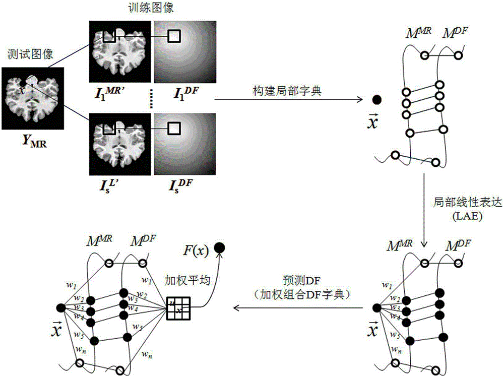
[0060] Ⅰ. The MR image block and the DF image block are located on two nonlinear manifolds, and any MR image block can be linearly expressed by its neighbor samples in the local space of the manifold;

[0061] Ⅱ. Under local constraints, the mapping from MR manifold to DF manifold is approximately a diffeomorphism mapping.

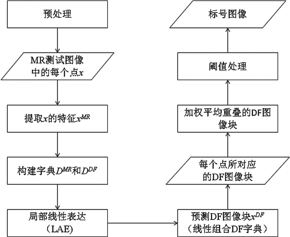
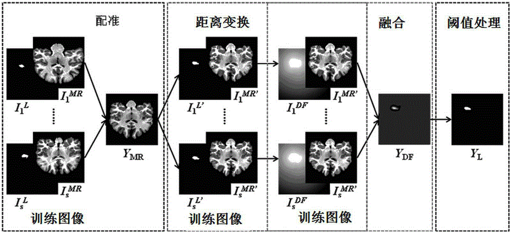
[0062] The MR image hippocampus segmentation method based on distance field fusion is carried out through the following steps:

[0063] (1) Normalize the initial MR test image to be segmented, remove the skull and offset field, and obtain the normalized MR test image. Specifically, the gray scale normalization method is used to normalize the MR test image, the BET algorithm is used to remove the skull, and the N4 algorithm is used to remove the offset field.

[0064] (2) Register the MR training set image in the pre-prepared train...

Embodiment 2

[0098] In order to verify the validity of the method of the present invention, the verification is carried out on the basis of 35 groups of MR brain data included in the database. The data of each set of MR brain data includes the T1-weighted MR images of the same patient and the corresponding labeled images of the hippocampus, among which 20 sets of data are randomly selected as the training set, and the remaining 15 sets of data are used as the test set. In the experiment, only the MR images of the subjects in the test set are used, and the labeled images of the hippocampus in the corresponding database are not used.

[0099] The present invention is based on the distance field fusion MR image hippocampus segmentation method, the method is based on two assumptions:

[0100] Ⅰ. The MR image block and the DF image block are located on two nonlinear manifolds, and any MR image block can be linearly expressed by its neighbor samples in the local space of the manifold;

[0101] ...


PUM


Abstract

The invention relates to a distance-field-fusion-based hippocampus segmentation method of an MR image. The method comprises: (1), normalization processing is carried out on an initial to-be-segmented MR test image and a skull and a biased field are removed, thereby obtaining an MR test image after normalization processing; (2), registering of an MR training set image and a training set label image to the MR test image is carried out; (3), distance conversion is carried out on the registered label image to obtain a distance field DF; (4), for a point X in the MR test image, an image block X<MR> is taken and is converted into a column vector; (5) a searching window is defined and an MR dictionary and a DF dictionary are selected; (6), the MR dictionary is used for expressing an MR test sample locally and linearly and a dictionary weight coefficient vector is solved; (7), a DF prediction image block vector of the test sample is obtained and is converted into an image block X <DF>; (8), the steps from (4) to (7) are repeated to obtain a DF value of each point; and (9), a label corresponding to each point in the test image is obtained. According to the invention, a target can be segmented accurately in an MR image.

Description

technical field [0001] The invention relates to the technical field of medical image analysis, in particular to a method for segmenting the hippocampus of an MR image based on distance field fusion. Background technique [0002] The hippocampus belongs to the gray matter structure of the midbrain. Hippocampus atrophy is a pathological manifestation of mental diseases such as Alzheimer's disease, schizophrenia, and depression. Clinically, these mental diseases can be diagnosed by analyzing the volume and shape of the hippocampus. Diagnosis. MRI can provide high-resolution anatomy, clear contrast, and a wide range of image sequences, which makes MRI technology popular in clinical diagnosis. Doctors can analyze the patient's brain structure through the patient's brain MR images, so as to make a diagnosis of the disease. Segmentation of the hippocampus in MR brain images is a prerequisite for hippocampus volume measurement and morphological analysis. Since the manual segmenta...

Claims


Application Information

Patent Timeline
IPC IPC(8): G06T7/00
CPCG06T2207/10088G06T2207/20021G06T2207/30016
Inventor 冯前进庞树茂阳维卢振泰
Owner SOUTHERN MEDICAL UNIVERSITY
Why Eureka
Try Eureka