
A detection kit for benign lesions and malignant lesions of oral mucosa and detection method thereof

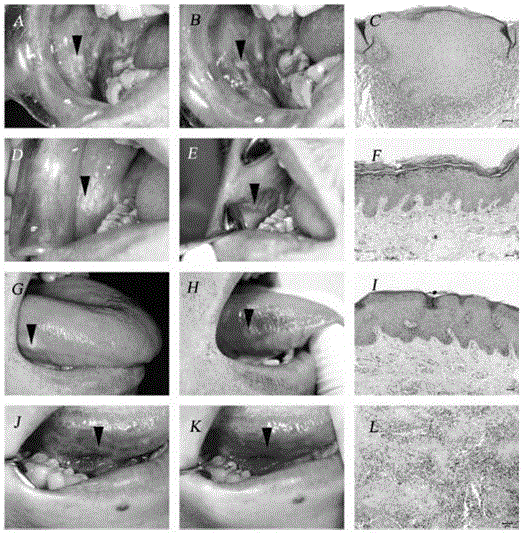
A detection kit and oral mucosa technology, which are applied in the field of benign lesions investigation and malignant lesion detection kits of oral mucosa, can solve the problems of promoting metastasis, affecting early diagnosis, and small oral lesions.

Active Publication Date: 2016-08-17
美泰克斯商贸(北京)有限公司
View PDF5 Cites 1 Cited by
  • Summary
  • Abstract
  • Description
  • Claims
  • Application Information

AI Technical Summary

Problems solved by technology

This method is direct and accurate, but because it is invasive, it may promote metastasis, and the oral lesion tissue is small, and false negatives may occur due to site deviation during sampling and sectioning. Therefore, this method is not easy to be introduced to patients early, which will affect early diagnosis.

Method used


Image

  • A detection kit for benign lesions and malignant lesions of oral mucosa and detection method thereof
  • A detection kit for benign lesions and malignant lesions of oral mucosa and detection method thereof
  • A detection kit for benign lesions and malignant lesions of oral mucosa and detection method thereof

Examples


Embodiment 1

[0034] (1). Oral inspection, record the details of the disease;

[0035] (2). Rinse mouth with clean water to remove food debris;

[0036] (3). Take the pre-check reagent and rinse for 1 to 5 minutes;

[0037] (4). Dip a cotton swab into the detection reagent 1 and rub it on the lesion area, and stain for 2-10 minutes;

[0038] (5). Take the inspection reagent 2 and rinse for 1 to 5 minutes;

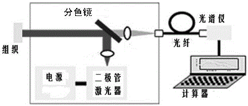
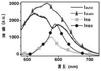
[0039] (6). Compare the colorimetric plate or auto-excited fluorescence detection instrument to semi-quantitatively or quantitatively evaluate the staining results.

[0040] The above-mentioned phosphate buffer is 0.01M, and the preparation method is as follows:

[0041] At a temperature of 0~4℃, weigh 7.9g NaCl, 0.2g KCl, 1.44g Na2HPO4 and 1.8g K 2 HPO4, dissolved in 800ml of distilled water, adjust the pH of the solution to 7.4 with HCl, and finally add deionized water to make the volume to 1L, and store in a refrigerator at 4°C for later use.

[0042] The preparation method of the above-mentioned ...

Embodiment 2

[0045] (1). Oral inspection, record the details of the disease;

[0046] (2). Rinse mouth with clean water to remove food debris;

[0047] (3). Take the pre-check reagent and rinse for 1 to 5 minutes;

[0048] (4). Dip a cotton swab into the detection reagent 1 and rub it on the lesion area, and stain for 2-10 minutes;

[0049] (5). Take the inspection reagent 2 and rinse for 1 to 5 minutes;

[0050] (6). Compare the colorimetric plate or auto-excited fluorescence detection instrument to semi-quantitatively or quantitatively evaluate the staining results.

[0051] The above-mentioned phosphate buffer is 0.01M, and the preparation method is as follows:

[0052] At a temperature of 0~4℃, weigh 7.9g NaCl, 0.2g KCl, 0.24g KH 2 PO4 and 1.8g K 2 HPO4, dissolve in 800ml distilled water, adjust the pH value of the solution to 7.4 with HCl, and finally add deionized water to make the volume to 1L, and store in a refrigerator at 4°C for later use;

[0053] The preparation method of the above-mention...


PUM


Abstract

The invention provides a kit for benign lesion identification and malignant lesion detection of the oral mucosa and a detection method thereof. The detection method comprises the following steps that 1, oral cavity examination is performed, and detailed information of lesions is recorded; 2, the mouth is rinsed with clean water to remove food debris; 3, a preexamination reagent is taken for gargling for 1-5 minutes; 4, a first examination reagent is taken in a dipping mode with a cotton swab to be embrocated on the partial lesions, and dyeing is performed for 2-10 minutes; 5, a second examination reagent is taken for gargling for 1-5 minutes; 6, semi-quantitative or quantitative evaluation is performed on the dyeing results by comparing the dyeing results with a color plate or a self-excitated fluorescence detecting instrument. The kit can be used for auxiliary differential diagnosis of the benign lesion and the malignant lesion of the oral mucosa and can assist a clinician to early diagnose an oral cavity cancer and monitor the malignant process of the oral precancerous lesions.

Description

Technical field [0001] The invention relates to a detection device kit, in particular to a detection kit for benign lesions and malignant lesions of the oral mucosa and a detection method thereof. Background technique [0002] Oral cancer refers to malignant tumors that occur in the oral cavity, including lip cancer, gum cancer, tongue cancer, soft and hard palate cancer, jaw bone cancer, mouth floor cancer, oropharyngeal cancer, salivary gland cancer, and maxillary sinus cancer, as well as facial skin Mucosal cancer, etc. Oral cancer is one of the more common malignant tumors of the head and neck. Currently, there are two types of diagnostic techniques used for oral cancer and precancerous lesions: [0003] 1. Traumatic technique: the histopathological method, the patient's diseased tissue needs to be excised, and then sliced ​​and observed under a microscope. This method is direct and accurate, but because it is traumatic, it has the possibility of promoting metastasis, and the ...

Claims


Application Information

Patent Timeline
Patent Type & Authority Patents(China)
IPC IPC(8): G01N1/30G01N21/25G01N21/64
Inventor 李成章陈汉正杜格非
Owner 美泰克斯商贸(北京)有限公司
Learn More