Three-dimensional hystero-salpingography imaging instrument

A technology of contrast imaging and fallopian tubes, which is applied in catheters, surgery, etc., can solve the problems of complex diagnostic methods and X-ray radiation, and achieve the effect of simple structure, small shape and convenient use

Inactive Publication Date: 2015-12-16
苏州斯科特医学影像科技有限公司
View PDF7 Cites 1 Cited by
  • Summary
  • Abstract
  • Description
  • Claims
  • Application Information

AI Technical Summary

Problems solved by technology

The traditional method requires obstetricians and radiologists to perform surgical examinations together. The diagnosis method is complicated, and there is X-ray radiation. The diagnosis time takes 2 to 3 days

Method used


Image

  • Three-dimensional hystero-salpingography imaging instrument
  • Three-dimensional hystero-salpingography imaging instrument

Examples


Embodiment Construction

[0012] The present invention will be further described below in conjunction with the drawings.

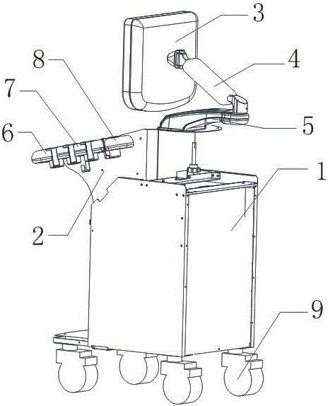
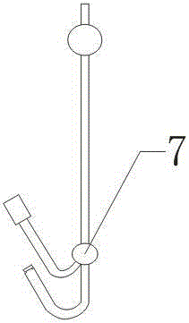
[0013] Three-dimensional hysterosalpingography imager, including host 1, PC end 2, display 3, bracket 4, support 5, transvaginal probe 6, contrast tube 7, spare probe 8, universal wheel 9, characterized by the setting of PC end 2 At the upper front part of the host 1, the display 3 is fixed on the support 5 through the bracket 4, and the support 5 is located above and behind the host 1, and the transvaginal probe 6, the radiography tube 7, and the spare probe 8 are arranged on the upper side of the host 1 in sequence. The transvaginal probe 6, the contrast tube 7, and the spare probe 8 are all connected to the host 1 through cables. The four corners of the host 1 are provided with universal wheels 9, and the tail part of the contrast tube 7 is the control end and the injection end. The transvaginal probe 6 and the spare probe 8 are three-dimensional ultrasound probes. The contrast tu...


PUM


Abstract

The invention relates to a three-dimensional hystero-salpingography imaging instrument. The three-dimensional hystero-salpingography imaging instrument comprises a host, a PC end, a displayer, a support, a supporting base, a per-vagina probe, a radiography tube, a standby probe and universal wheels. In actual usage, an obstetrician extends the front end of the radiography tube into the uterus through the control end at the tail end of the radiography tube in combination with the per-vagina probe, then directly injects perfluoropropane-albumin mincrosphere injection ultrasonic contrast agents in a diagnosis room through an injection end and then places the per-vagina probe or the standby probe at the bottom of the vagina, and after injection radiography, the filling and smoothing conditions of the uterus and uterine tubes are observed in real time. Because the selected perfluoropropane-albumin mincrosphere injection ultrasonic contrast agents are water-soluble contrast agents and can be dispersed inside the pelvic cavity in real time, the smoothing and filling conditions of the uterus and uterine tubes are convenient to diagnose, and the purpose of diagnosis is achieved.

Description

Technical field [0001] The invention relates to ultrasonic diagnosis and treatment equipment, in particular to a three-dimensional hysterosalpingography imaging instrument. Background technique [0002] Ultrasound diagnosis mainly uses the good directivity of ultrasound and the physical characteristics of reflection, scattering, attenuation and Doppler effect similar to light. It uses its different physical parameters, uses different types of ultrasound diagnostic equipment, and uses various scanning methods. , Transmit ultrasound into the human body and propagate in the tissues. When the acoustic impedance of normal tissues or pathological tissues is different, the interface formed by them will reflect and scatter, and then receive this echo signal and perform detection and other processing After that, it is displayed as a waveform, curve, or image. [0003] As a kind of ultrasound diagnosis, B-ultrasound can clearly display various cross-sectional images of various organs and su...

Claims


Application Information

Patent Timeline
IPC IPC(8): A61B8/12
Inventor 高兴斌周正帮黄孟钦吴丽丽
Owner 苏州斯科特医学影像科技有限公司
Why Eureka
Try Eureka